Last Updated on August 2, 2019
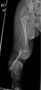
Absence of tibia is a rare birth defect characterized by a deficiency of the tibia while the other bones of the lower leg relatively intact. The deficiency may occur in one or both legs. The right side is more commonly affected [72%]. Bilateral occurrence is found in about 30%.
The absence of tibia could be an isolated birth defect but is commonly part of part of a recognized syndrome such as Werner’s syndrome, tibial hemimelia-polysyndactyly-triphalangeal thumb syndrome, and CHARGE syndrome.
Most isolated cases occur sporadically in people with no family history.
The underlying cause is generally unknown.
There is a high-risk association of congenital dislocation of the hip, aphalangism of upper and lower limbs, proximal femoral focal deficiency and hypoplasia of the femoral condyles.
There are varying degrees of tibial absence.
Presentation in Congenital Absence of Tibia
Tibial hemimelia presents as a shortened leg with knee and ankle deformities. Varus in the ankle and a knee flexion contracture in the knee are can occur. There is a deficiency of collateral ligaments as well and this leads to instability of the joints due to a lack of collateral ligaments.
The patellar bone may be absent and so could be quadriceps muscle. When present, their function is reduced.
The tibia may be shortened, dysplastic, or absent. The tibia may also present as an anlage—a fibrous remnant that does not appear on x-rays.
The fibula is normal or dysplastic. It is often dislocated from the knee. Foot deformities are present as well.
The spectrum of the tibial deficiency ranges from a congenitally short tibia with relative fibular overgrowth to complete absence of the tibia
Differential Diagnoses
The absence of tibia can be distinguished from fibular hemimelia and congenital deficiency of knee.
[foot and ankle are always in varus with the absence of tibia and almost always in valgus in fibular hemimelia.]
Imaging

Radiographs recommended are anteroposterior and lateral views of leg showing complete tibia fibula. Joints on either side [knee and ankle] should be included in the x-ray.
X-rays show a deficiency of the tibia. Early radiographs may show small and minimally ossified distal femoral epiphysis
Treatment of Congenital Absence of Tibia
Treatment is surgical. Following procedures could be done depending on the condition of absence of tibia.
Knee Disarticulation Followed by Prosthetic Fitting
The limb is amputated at the knee and when ready a prosthesis is fit. The procedure is indicated in the complete absence of the tibia and when there is no active knee extension present
Tibiofibular Synostosis with Modified Syme Amputation
In this procedure, the available proximal tibial stump is fixed to the fibula. Syme amputation is an amputation done through the ankle joint but the heel pad is saved.
The procedure is indicated when proximal tibia present with an intact extensor mechanism and there is minimal flexion contracture.
Syme amputation is needed because of ankle diastasis and foot deformities making them unusable for locomotion.
Brown Procedure (centralization of fibula under femur) is no longer recommended due to the high failure rate
Dror Paley uses a different classification and different approach to treatment to maximize limb saving. Validity, reproducibility, and acceptance of his approach by standard literature are yet to be seen.