• Skip to main content
  • Skip to primary sidebar
  • Skip to footer
  • General Ortho
  • Procedures
  • Spine
  • Upper Limb
  • Lower Limb
  • Pain
  • Trauma
  • Tumors
  • Newsletter/Updates
  • About Us
  • Contact Us

Bone and Spine

Orthopedic health, conditions and treatment

Cortical Bone and Cancellous Bone

By Dr Arun Pal Singh

In this article
    • What is Cortical Bone
    • Cancellous Bone or Spongy Bone

Cortical bone and cancellous bone are two types of osseous tissues of the human skeleton which differ in their structure and distribution. The difference in their structures and distribution is designed to carry different functions.

What is Cortical Bone

Cortical bone refers to the thick outer surface of typically a long bone [for example humerus or femur shaft] that ensheathes the cavity of the bone called medulla. The sheath or outer shell formed by the cortical bone is called cortex.  Cortical bone is also called compact or lamellar bone and provides strength to all the long bones of the body, for example, femur.

Cortical bone contributes about 80% of the weight of a human skeleton.  It is much denser than cancellous bone,  harder, stronger and stiffer.

At the microscopic level, the structural arrangement of a cortical bone is different than cancellous. Compact bone consists of closely packed osteons or Haversian systems. The osteon consists of a central canal called the osteogenic (Haversian) canal, which is surrounded by concentric rings (lamellae) of the matrix.

[See diagram below]

Between the rings of the matrix, the bone cells (osteocytes) are located in spaces called lacunae. Small channels (canaliculi) radiate from the lacunae to the osteonic (Haversian) canal to provide passageways through the hard matrix.

The Haversian systems are packed tightly together to form what appears to be a solid mass. The osteonic canals contain blood vessels that are parallel to the long axis of the bone. These blood vessels interconnect, by way of perforating canals, with vessels on the surface of the bone.

[Read more on Anatomy and Physiology of bone]

Structural Arrangement of Cortical Bone and Cancellous Bone
Structural Arrangement of Cortical and Cancellous Bone

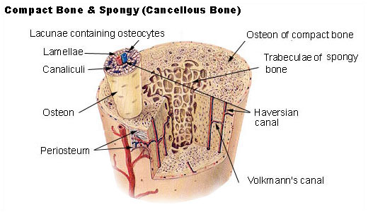
Image Credit: Public domain image, from Wikipedia

Cortical bone is arranged for facilitating the following functions

  • To support the whole body weight
  • Protect organs
  • Provide levers for movement
  • Store and release calcium.

Cancellous Bone or Spongy Bone

Cancellous bone is also called trabecular bone or spongy bone.

Compared to compact bone, which is the other type of osseous tissue, it has a higher surface area but is less dense, softer, weaker, and less stiff.

Cancellous bone
Cancellous Bone
Image Credit:OERPUB

It typically occurs at the ends of long bones, proximal to joints and within the interior of vertebrae.

For example, distal end radius, proximal humerus or proximal femur, all are examples of cancellous bone.

Microscopically, cancellous bone consists of plates (trabeculae) and bars of bone adjacent to small, irregular cavities that contain red bone marrow.

The canaliculi connect to the adjacent cavities, instead of a central Haversian canal, to receive their blood supply. It may appear that the trabeculae are arranged in a haphazard manner, but they are organized to provide maximum strength similar to braces that are used to support a building.

The trabeculae of spongy bone follow the lines of stress and can realign if the direction of stress changes.

Cancellous bone has a greater surface area and is ideal for metabolic activity e.g. exchange of calcium ions.

In osteoporosis, cancellous bone is more severely affected than cortical bone.

Cancellous bone is highly vascular and frequently contains red bone marrow where hematopoiesis, the production of blood cells, occurs. Bone marrow also acts as a source of stem cells.

All the procedures for the study of bone marrow are done in spongy bone

Both cortical and cancellous bones together form the skeleton and serve their respective functions.

Spread the Knowledge
  • 13
    Shares
  •  
    13
    Shares
  • 1
  • 12
  •  
  •  
  •  

Filed Under: Anatomy

About Dr Arun Pal Singh

Arun Pal Singh is an orthopedic and trauma surgeon, founder and chief editor of this website. He works in Kanwar Bone and Spine Clinic, Dasuya, Hoshiarpur, Punjab.

This website is an effort to educate and support people and medical personnel on orthopedic issues and musculoskeletal health.

You can follow him on Facebook, Linkedin and Twitter

Reader Interactions

Comments

  1. Injuries & Conditions says

    January 10, 2013 at 12:48 pm

    Bone is a important thing on our body parts as I think That is why we need know about bone as well. In this post you’ve shared some important info about the Types of Bone – Cortical and Cancellous as well. This is really effective to all. Thanks!!!

  2. Bhagyashree says

    September 16, 2017 at 9:37 am

    Thanks a lot. Helped me so much to understand the difference between the two. :)

  3. Arwa AbouGhareeb says

    September 22, 2017 at 10:35 pm

    Great job summarizing and comparing the two types of bone tissue. I’m just unsure about the last paragraph which states that cancellous bone is highly vascularized, as spongy bone does not contain blood vessels.

  4. Dr Arun Pal Singh says

    September 25, 2017 at 11:38 am

    I am glad Bhagyashree it helped you.

  5. Dr Arun Pal Singh says

    September 27, 2017 at 4:10 pm

    Thanks for the appreciation. Cancellous bone is very vascular. It has to be because it is the place where blood cells are formed by marrow.

  6. hassan says

    November 27, 2017 at 4:09 pm

    any one help me my mother left leg is bone cortices are intact

  7. Lars Edwards says

    February 17, 2018 at 1:52 am

    Would cortical and cancellous bone explain color variation on an MRI? Black vs very light gray? Or would light gray almost white coloring be of concern on an MRI?

  8. Dr Arun Pal Singh says

    February 18, 2018 at 7:11 pm

    The cortices are blacker, medulla almost looks similar. The concern should arise if the difference is focal rather than widespread. If you are able to put across your specific concern, I would be in better position to reply.

  9. Lars Edwards says

    February 18, 2018 at 9:12 pm

    My 10 year old daughter broke her tarsal bone in her foot. It got hit with a bowling ball ( significant force). This is her 3rd broken bone. X Ray was questionable so they ordered an MRI. It showed the break in much better detail. The doctor was concerned about the color contrast from the bottom of the tibia (dark) to the tarsal (white or light grey). From my best recollection……
    I wish I had a second look at the MRI because then I would have measured certainty of what I’m relating to you. But for discussion sake, let’s pretend that’s the exact concern of the bones mentioned. I do recall him saying possibly osteomalasia? He ordered blood work as precaution, vitamin D and calcium, all minerals were nominal. So what could be left or overlooked? I’ll get better information if necessary. Thanks so much for your input!

  10. Dr Arun Pal Singh says

    February 20, 2018 at 11:19 am

    Lars Edwards,

    I understand that you are concerned about something doctor said but there are too many ifs and buts to answer it in proper way. But I do understand that sometimes incidental findings could also be significant.

    I think the best way is to discuss it with the treating doctor, understand the concerns raised and their implications. If there is an MRI finding that bothers, review of the image from radiologist can be asked.

    Osteomalacia as term is used in adults. In children, the similar condition is called rickets[some authors do call it pediatric osteomalacia]. In rickets, the bone keeps growing at the end but the calcification is delayed. But I doubt that could cause of darker bone. [Its further confusing that MRI has two kinds of images T1 and T2 weighted and I do not even know what you may be referring to]

    Please talk to your doctor and ask it is wiser to investigate further or okay to wait and watch.

    Take care.

  11. Evanton ville says

    September 9, 2019 at 2:04 pm

    Which bone heals fast is it cortical or cancellous and give me reasons

  12. Arun Pal Singh says

    September 15, 2019 at 9:00 am

    Evanton,

    Cancellous bone because of the surface area of contact is more and the number of active bone cells participating in the healing process is higher.

Leave a Reply Cancel reply

Your email address will not be published. Required fields are marked *

This site uses Akismet to reduce spam. Learn how your comment data is processed.

Primary Sidebar

Types of lesion in tuberculosis of spine

Tuberculosis of Spine Presentation and Treatment

Tuberculosis of spine or TB spine or spinal TB was first described by Percivall Pott, after whom it is also referred to as Pott’s disease or Pott’s spine. Tuberculosis of spine is the commonest form of skeletal tuberculosis. It constitutes about 50 percent of all cases of bone and joint TB. There are few other […]

Tuberculosis of foot, calcaneum osteolytic variety

Tuberculosis of Foot

Tuberculosis of foot is rare and constitutes less than 10% of cases of osteoarticular tuberculosis. Calcaneum is the most common bone involved followed by subtalar joint and midtarsal joints. Pathophysiology Bonewise, the order of involvement in decreasing order is calcaneum, talus, first metatarsal, navicular, first and second cuneiforms cuboid and others. The infection first lodges […]

fracture-proximal-phalanx-hand

Phalanx Fractures of Hand [The Broken Fingers]

Phalanx fractures are common hand injuries. Phalanges are small long bones of the hand that make our fingers and thumb. A similar arrangement of bones is also found in toes and those bones are also called phalanges. Each finger is formed by three phalanges. Proximal phalanx Middle phalanx Distal phalanx Thumb has only two phalanges, […]

Representative Image of spondylosis

Spondylosis – Definition, Causes and Treatment

Spondylosis as a medical term denotes degenerative changes that occur in the spine as a natural aging process for some other reason. The term degeneration means the wear and tear that accumulates. The bone spurs and degeneration of intervertebral discs are examples of degeneration. The term is from the Ancient Greek word ‘spondylos’ which [plural […]

scapulothoracic bursitis bursa locations

Scapulothoracic Bursitis Causes and Treament

Scapulothoracic bursitis is inflammation of the scapulothoracic bursae present between the scapula and rib cage. Symptomatic scapulothoracic bursitis is not very common. Another similar but different term used is scapulothoracic crepitus [snapping scapula] and it is important to highlight the difference. Scapulothoracic crepitus is referred to as thumping or popping sounds that occur with scapulothoracic […]

ankle arthrocentesis sites

Ankle Arthrocentesis or Ankle Joint Aspiration

Ankle arthrocentesis like other joint aspirations could be diagnostic or therapeutic. A good knowledge of ankle joint helps to perform the arthrocentesis more efficiently and safely. Relevant Anatomy of Ankle Joint The ankle joint is a hinged synovial joint formed by talar bone, tibia, and fibula. Plantar flexion and dorsiflexion are the main movements of […]

Bertolotti syndrome is caused by lumbosacral transitional vertebra.

Bertolotti Syndrome – Causes and Treatment

Bertolotti syndrome is the presence of a variation of the fifth lumbar vertebra with a large transverse process, either articulated or fused with the sacral basis or iliac crest, producing a chronic, persistent lower back pain. It may also cause isolated L4/5 disc disease. Bertolotti described a unilateral and bilateral enlargement of the transverse process […]

Browse Articles

Footer

Pages

  • About
    • Policies
    • Contact Us

Featured Article

Acromioclavicular Joint Injuries

Acromioclavicular joint injuries are also known as shoulder separations, acromioclavicular joint separation or acromioclavicular joint dislocation and … [Read More...] about Acromioclavicular Joint Injuries

Search Articles

© Copyright: BoneAndSpine.com