Last Updated on August 2, 2019
An external fixator is a useful tool in the management of fractures and certain difficult orthopedic problems such as limb length discrepancy. External fixation a useful tool in fracture management and in the case of pelvic fracture it may be a primary life-saving device
With external fixation, pins and/or wires are percutaneously inserted into the bone and held in place by an external frame.
[Know more about other types of fixations]
External fixation is most successful in superficial bones like tibia than deeper bones like femur or humerus – here the chance of pin tract sepsis is greater.
External fixators consist of modular components which are assembled to form a stable construct between bone fragments and an adjustable beam system. The beam system is joined to the bone by means of a number of pins screwed into the bone.
The technique of external fixation was popularized in the mid-20th century by Hoffman.
Indications for Use of External Fixators
- Limb length discrepancy surgeries
- Arthrodesis
- Correction of angulatory or rotator deformity
- Bone segment transportation to fill the bone gap
- Temporary fixation of open fractures
- Maintains stability
- Eases dressing
- When definitive surgery is delayed for some reason
- Rapid Stabilization
- Pelvic fixation to stop bleeding
- Rapid fixation of polytrauma patients
- Definitive external fixation of fractures especially intraarticular fractures
- Ligamentotaxis
- To position limb in the desired position as in nerve or tendon repair
- Infected fractures
- Burns
Advantages of External Fixation
- Provides rigid fixation when other forms of immobilization are not feasible
- For example severe open fractures
- Allows compression, neutralization, or fixed distraction of the fracture fragments.
- Allows limb surveillance and wound care without disturbing alignment [compare with casting]
- Dressing changes
- Skin grafting
- Bone grafting
- Frequent debridement and irrigation
- Allows joint mobilization
- Allows limb elevation by suspending frame if needed
- Allows early patient ambulation
- External fixators cause less disruption of the soft tissues, osseous blood supply, and periosteum.
Disadvantages of external fixation
- Risk of pin tract infection
- Fracture through pin tracts after frame removal.
- Cumbersome and needs meticulous care.
- Joint stiffness when the joint is immobilized
Types of External Fixators
In strictest sense, there are two types of fixators – unilateral and circular. A combination of two is called hybrid fixators.
Unilateral Fixator [Modular AO Type]
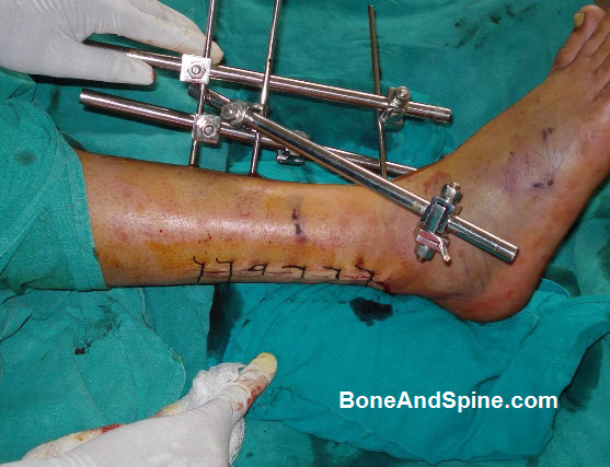

They are called so because they are generally are distinguished from circular frames in that they are positioned on one side of the limb. Unilateral frames allow the limb to remain functional, avoid complications, and provide bony stability
Two most common designs are the bulkier
Monobody designs
The monobody frames have considerable intrinsic stability owing to their heavy and rigid design.
Pin-to-bar fixators.
These are kind of fixators use a combination of Schanz screws, rods and clamps which are assembled to form a construct.
Circular Fixators
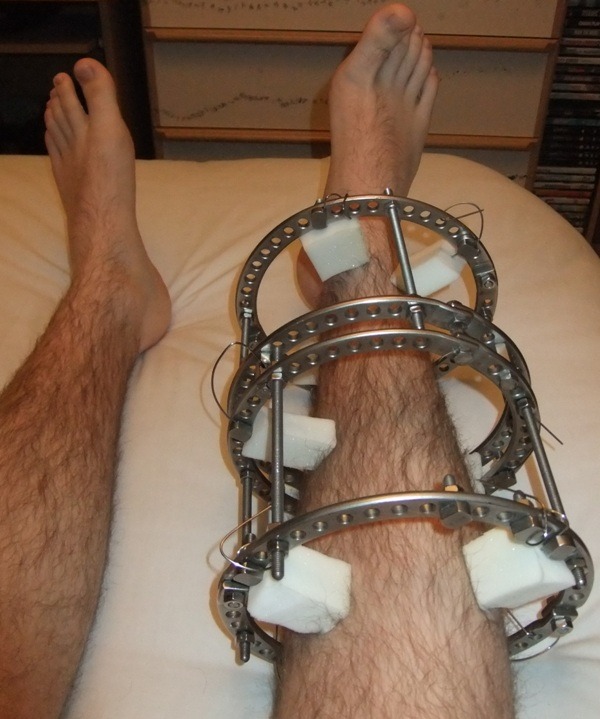
Image Credit: Wikipedia
These kinds of external fixators use construct formed by circular rings, wires, connecting rods, and struts. This is quite a versatile type of external fixator. A partial ring is commonly used around the proximal and around the shoulder and proximal femur where a full ring would not fit comfortably.
Unilateral or Modular AO External Fixator Assembly
Components of this fixator are given below.
- Schanz Screws
- Connecting rods
- Clamps
Each of the components can come in different dimensions to suit the scale of the bone to which they are to be applied and to permit variation in the shape and configuration of the final bone-external fixator construct.
Schanz screws
Schanz screws are partially threaded pins. These are available in different diameters and lengths of the shaft and threaded part and with different tips.
Standard screws have trocar-shaped tips. They require pre-drilling. Self-drilling and self-cutting screws are available.
Schanz screws are available in steel and titanium. Shanz pins with hydroxyapatite coating are also available. This makes bone purchase better and allows easier osseointegration, preventing loosening. Pins with hydroxyapatite coating may be preferred for long-term application of external fixators, eg, bone transport or deformity correction.
Rods/tubes
The AO fixators consist of systems in four sizes, depending on the size of the rod:
- Large: 11 mm tubes/rods with Schanz screws from 4 to 6 mm;
- Medium: 8 mm tubes/rods with Schanz screws from 3 to 6 mm;
- Small: 4 mm tubes/rods with Schanz screws from 1.8 to 4 mm;
- Mini: 2 mm system for fingers. Includes multipin clamps for K-wires and 2 mm longitudinal rods.
All systems are compatible with each other.
The large 11 mm system contains both steel tubes and carbon fiber rods.
Pre-curved contoured and T shaped rods are also available.
Currently, the 4.7mm short threaded Schanz screw is used in cortical bone and the 5.0mm long threaded screw in the cancellous bone. For hand and wrist application smaller sizes are used.
Connecting rods are made of stainless steel of carbon fiber. The latter are very strong and are also radiolucent which is helpful when assessing bone alignment on x-ray.
Clamps
The clamps provide the connection between the tubes or rods and the pins. Likewise, rods or tubes can be connected to each other using the appropriate clamps (tube-to-tube). If one clamp allows the connection of both tubes and rods, they are called combination clamps.
Both single-pin and multipin clamps are available.
Clamps are available in three sizes with identical clamp design and application technique.
A larger tube to tube clamp permits two fracture components to be held together in relatively stable alignment irrespective of the position the Schanz pins take up in the bone.
Stability of Unilateral or Modular AO External Fixator
The bending stiffness of unilateral fixators is dependent on the plane of the half pins and the plane of loading with anteriorly mounted frames providing greater bending stiffness. When these frames were loaded out of the plane, with varus–valgus and torsional forces, they had poor control of the bone fragments with significant motion at the fracture site.
More stability would be obtained from a multiplanar system.
The stiffness of the frame depends upon the following factors:
- Closer the Schanz screws to the fracture, stiffer the construct
- Farther the last Schanz screw placed on the fragment on each side of the fracture, stiffer the construct
- Closer the longitudinal connecting tube/bar to the bone. Stiffer the construct
- Two bars/tubes are stiffer than one;
- Frame configuration – The stiffness of the construct depends on the assembly. Different constructions of tubular external fixator which will produce increasing levels of stability are
- Unilateral uniplanar single-tube fixator.
- Unilateral uniplanar single-tube modular fixator.
- Unilateral uniplanar double tube fixator.
- Unilateral biplanar frame (delta-frame or triangular frame).
- Bilateral frame with transfixing pins [Hardly used now].
Circular Fixator
Ilizarov fixator is a typical circular fixator. Circular fixators consist of a series of rings or arches which are connected to each other by connecting rods and rings are fixed to the bones by means of tensioned wires.
Many modifications can be added and accordingly the implant used in particular fracture is used according to the indication for use and goals of the surgery.
Ilizarov fixator is very versatile fixator and most people know it because it is used for lengthening the bones.
Ilizarov external fixator is discussed in detail separately.
In circular external fixators, frame stability is greatly impacted by ring properties. Smaller diameter rings are more stable than larger rings of the same thickness but a ring should not put pressure on the tissues and the final size is dictated by limb girth.
Different diameter rings may be used in the same frame to adjust to contours of the limb. Centralizing the bone is preferred but eccentric positioning has been found to have no adverse effect.
The closer the rings are, the better is the frame stability.
The stability of the construct is increased by
- Use of a greater number of rings
- Shorter distances between rings
- Increasing the span of the rings across the bone controlling both near and far ends of each segment
- Increasing the number of connections between the rings
- Increasing number of points of fixation to the bone.
Best Practice Tips for External Fixator
- Placing pins with aseptic technique
- Protecting soft tissues so that no necrotic tissue is left to encourage infection
- Drilling a pilot hole to remove bone debris and to reduce frictional resistance and so heat production during pin insertion.
- Achieving a firm pin fit with a radial distribution of forces – the pin is 0.2mm larger than the pilot hole and this induces compression on the bone termed radial pre-load.
Complications of External Fixation
Complications may arise in the fixator itself or most commonly at the bone-pin interface. As with any fracture especially in severe injury complications may occur at the fracture as a direct result of the injury.
Pin Tract infection
This may be the most common complication, occurring in about 30% of patients. The infection varies from minor skin inflammation to osteomyelitis requiring sequestrectomy.
Pin site infection occurs more when
- Pin is inserted at a place with greater soft tissue
- Skin tethering over the pin
- Inadequate pin care
Neurovascular Injury
The radial nerve in the distal half of the arm and proximal half of the forearm, the dorsal sensory radial nerve just above the wrist, and the anterior tibial artery and deep peroneal nerve at the junction of the third and fourth quarters of the leg are the structures most often involved.
The pins are known to penetrate vessels, cause thrombosis when they are adjoining the vessel, cause late erosion of the vessel, arteriovenous fistulas, and the formation of aneurysms.
Muscle Fibrosis and Tendon Rupture
Pins inserted through tendons may restrain normal excursion and can lead to tendon rupture, or muscle fibrosis
Fracture Complications
Nonunion and delayed union can be seen with any mode of fixation and can occur with external fixator also.
Refracture can occur after fixator removal and the fracture needs protection for an extended period after fixator is removed.