Last Updated on November 22, 2023
The femoral anteversion refers to the normal rotation of the femoral neck in relation to the femoral shaft. It is a normal anatomical occurrence when it occurs within the normal range. However, in some persons, for one reason or other the femoral anteversion value or the rotaion of the neck with respect to rest of the femur is more than the normal range.
This is called increased or exaggerated femoral anteversion and can lead to various problems as we would discuss below.
Many articles refer to both as ‘femoral anteversion’ only but to avoid confusion, it is best to have clarity of the issue.
Femoral Anteversion and Use of the Term
Before we move further on, it is better to understand what is anteversion and retroversion of an organ.
Anteversion and Retroversion describe the relative rotation of an organ or part of it.
- Anteversion means rotated forwards (towards the front of the body)
- Retroversion means rotated backward (towards the back of the body)
The version is in comparison to a reference position.
For example, the normal uterus is typically slightly anteverted.
In musculoskeletal science, the terms are important in the hip and shoulder.
For example, in the hip, the neck of the femur is anteverted slightly. This is termed as femoral anteversion and is part of the normal anatomy of the neck of the femur.
The femoral head has 12 to 15º of anteversion to the line connecting the posterior aspect of both femoral condyles.
Similarly, the normal humeral head has 30º of retroversion to the frontal axis of the elbow joint.
An increase in femoral anteversion more than the typical value range can cause problems in the alignment and gait and is an abnormality.
Thus, femoral anteversion is normal within a certain range. But an exaggerated level is an abnormality.
Thus technically speaking there is normal anteversion of the femur and there is abnormal anteversion of femur [when the normally present anteversion is exaggerated.]
However, in literature and in communication, both are addressed as femoral anteversion. In fact, I have come across various articles that relate the term femoral anteversion as an abnormality only.
Normally, the acetabulum in a similar manner is slightly anteverted.
Knowing the right version is important in designing arthroplasty implants and placement during surgery.
Thus the femoral neck is normally anteverted with respect to the rest of the femur [see the diagram]. It is considered abnormal only if it significantly differs from the average value of a patient of the same age.
Note: When we say that the femoral neck is anteverted, we are taking it to the rest of the femur for comparison. this is because the neck of the femur is not in the same plane as the shaft of the femur but rather anteriorly lifted or rotated when compared to that plane.
Change in Femoral Anteversion Angle with Age
- Newborn – 31 degrees
- 5 years – 26 degrees
- 9 years – 21 degrees
- 16 years – 15 degrees by 16 years of age.
Definition
Femoral anteversion is defined as the angle between an imaginary transverse line that runs medially to laterally through the knee joint and an imaginary transverse line passing through the center of the femoral head and neck.
Normal femoral anteversion in adults is 15 and 20 degrees from the frontal plane of the body.
The term medial femoral torsion is also used to describe femoral neck anteversion and is thought to result from medial or internal rotation of the limb bud in early intrauterine life.

Abnormal or Excessive Femoral Anteversion
An excessive femoral anteversion is a clinical problem. Some people refer to it simply as femoral anteversion.
It is a congenital condition where there is an increased anteversion of the femoral neck relative to the femur. It is caused often by intrauterine positioning.
The diagnosis is made clinically by the presence of –
- Intoeing – The feet is turned inwards when the child walks or runs [also called pigeon-toed.]
- Intoeing can be seen in younger children in metataesus adductus and tibial torsion in younger children. Intoeing after 3 years of age is usually due to increased anteversion of femur.
- There is an increased internal rotation of the hip along [with decrease in external rotation]
The condition is often bilateral with girls being affected more than boys.
Abnormal femoral anteversion is a developmental abnormality. The normal child is born with 31-40 degrees of femoral anteversion. This gradually decreases to 10 to 15 degrees at adolescence and generally improves with further growth.
Often, the intoeing resolves as the child grows, by 10years of age. In few cases surgical correction may be warranted
Clinical Presentation and Physical Examination
The usual complaint is of in-toeing gait in early childhood.
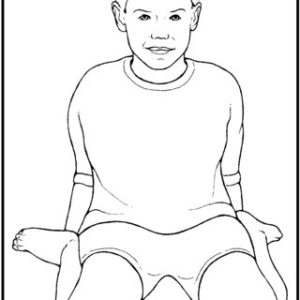
The child may sit in a unique posture or W position.

Determination and Significance of Femoral Neck Anteversion – Courtesy: Researchgate [accessed 18 Jun, 2023]
In extreme cases, sports activities and activities of daily life may be affected in older children.
There may be complaints of frequent tripping during walking.
On examination, the hip is found to have increased internal rotation and decreased external rotation.
The patella is rotated inwards when walking [differentiates from other causes of in-toeing like metatarsus adductus and tibial torsion]
Craig’s test measures the clinical value of the degree of femoral anteversion. This test is also called Trochanteric angle prominence test.
Craig’s Test
Imaging is not commonly required. However, CT can be used for the exact measurement of anteversion in selected cases.
This involves 3 images or scans—2 proximal and 1 distal. One image defines the location of the center of the femoral head, the second image defines the base of the femoral neck, and the third image defines the distal femoral condylar axis.
The angle in the transverse plane between the intersection of the plane of the neck and the condylar plane defines the angle of anteversion.
Treatment of anteversion of the neck of the femur mostly requires observation as most of the cases outgrow as age increases. This is because there is a decrease in the anteversion as the child grows.
Some patients would require derotational femoral osteotomy for surgical correction. It is a type of intertrochanteric osteotomy. This is indicated when there is less than 10 degrees of external rotation on examination in a child who is older than 10 years of age.
Femoral Retroversion or Retroversion of Hip
When the femoral neck angle is less than the average range [ 15-20 degrees], it is called femoral retroversion or hip retroversion.
If the anteversion is less than the normal average or the inclination of the femoral neck is in the opposite direction. Femoral neck retroversion is present when the head and neck of the femur are angled less than the average anteversion angle along the frontal plane of the body.
In some cases, the femoral head and neck may even be angled backward from the frontal plane of the body.
Patients who had decreased femoral neck anteversion angle angles also were more likely to have osteoarthritis of the hip.
References
-
Fabry G, Cheng LX, Molenaers G. Normal and abnormal torsional development in children. Clin Orthop Relat Res. 1994 May;(302):22-6.Sass P, Hassan G. Lower extremity abnormalities in children. Am Fam Physician. 2003 Aug 1;68(3):461-8. Erratum in: Am Fam Physician. 2004 Mar 1;69(5):1049. PMID.