Last Updated on October 28, 2020
Flexor tendon pulley system consists of annular ligaments of the fingers or A pulleys, and cruciate pulleys [C pulleys].
Flexor pulley system consists of following
- Palmar Aponeurosis pulley
- 5 Annular pulleys
- 3 Cruciform pulleys.
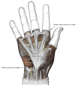
Together, these form a fibro-osseous tunnel on the palmar aspect of the hand through which passes the deep and superficial flexor tendons.
Flexor tendon pulley system maintains flexor tendons close to joint’s axis of motion and prevents bowstringing.
Digital flexor sheath is a synovial sheath which consists of membranous and retinacular parts.
Membranous part is composed of visceral and parietal layers. It carries the flexor digitorum profundus and flexor digitorum superficialis tendons in the distal aspect of the hand.
The flexor pulley system is formed of the retinacular component which condensates and are arranged in cruciform, annular pulleys and transverse patterns. These structures overlie the membranous, or synovial, lining.
Digital sheath serves following functions
- Facilitates smooth gliding of the tendons
- Pulleys from retinacular component provide a mechanical advantage to flexion
- Synovial fluid bathes and provides nutrition.
Components Flexor Tendon Pulley System
Palmar Aponeurosis Pulley
It is formed of transverse fascicular bands approximately 1 cm in width arising from the palmar aponeurosis.
The proximal edge of the pulley is 1-3 mm proximal to the origin of the membranous tendon sheath and the distal edge is approximately 8-10 mm from the proximal edge of the first annular pulley.
A vertical septum anchors this pulley to the deep transverse metacarpal ligament beneath the tendons. This pulley moves closer to tendon surface during grasping due to increased tension on the palmar fascia by the flexor carpi ulnaris and palmaris longus muscles.
Annular Pulleys
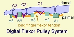
A1 Pulley
A1 pulley is first annular pulley which arises from the palmar plate and proximal portion of the proximal phalanx. It is at the level of metacarpophalangeal joints and is approximately 8 mm in width.
The proximal edge of the 1st annular pulley lies about 2 cm from the proximal finger crease and the distal edge of A1 pulley lies about 1 cm from the proximal finger crease.
At the level of the A1 pulley, the flexor digitorum superficialis bifurcates and allows flexor digitorum profundus to pass superficial..
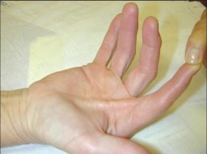
This pulley is frequently involved in trigger finger.
A2 Pulley
The second annular pulley originates from periosteum of the proximal and lateral areas of the proximal phalanx and is approximately 20 mm in width. This pulley consists of oblique fibers and is of all of the flexor tendon sheaths, this is probably the most important.
A3 Pulley
This is the third annular pulley and is located at the level of the proximal interphalangeal joint. It arises from the volar palmar plate and is approximately 3 mm in width.
A4 Pulley
A4 pulley arises from periosteum of mid-portion of the middle phalanx. It is similar to A4 pulley that oblique fibers overly annular fibers. This pulley is also to be preserved during surgery.
It is about 7 mm in width. It is considered the second most important flexor tendon sheath pulley (after the A2) and the most important biomechanical pulley for maintaining independent interphalangeal joint function.
A5 Pulley
This pulley is located proximal to distal interphalangeal joint,, just proximal to the termination of the membranous sheath. With a width of 4 mm, the A5 pulley is the thinnest of the 5 annular pulleys and has a width of 4 mm.
Cruciform pulleys
Also called cruciate pulleys, these are three pulleys that function to prevent sheath collapse and expansion during digital movements.
C1 Pulley
This cruciate pulley lies just distal to the A2 pulley
C2 Pulley
This pulley is located in the space between the A3 and A4 pulleys
C3 Pulley
This is located distal to the A4 pulley, though a number of variations have been described.
Flexor Tendon Pulley System of Thumb

Thumb has a separate flexor tendon pulley system.
The flexor sheath of thumb originates proximal to the radial styloid in the wrist and carries the single flexor pollicis longus tendon.
The retinacular system consists of 3 separate pulleys overlying the membranous sheath. These are
A1 Pulley
The first annular pulley is located at the level of the metacarpophalangeal joint. It is approximately 1 cm wide and originates from the volar plate and base of the proximal phalanx.
Oblique Pulley
This pulley overlies the flexor sheath at the midportion of the proximal phalanx. Oblique pulley originates at proximal half of proximal phalanx is about 10 mm in length, and blends with a portion of the adductor pollicis insertion.
It is most important pulley in thumb which facilitates full excursion of flexor pollicis longus and prevents bowstringing of flexor pollicis longus.
A2 Pulley
The second annular pulley is 1 cm in width and attaches to the volar plate of the interphalangeal joint.
Bowstring Phenomenon

To understand bowstring we need to imagine bow and string where string is straight and bow is is in smooth convex fashion.
But when we flex our finger, the different finger joints are at different angles. This is made possible by pulleys which keep the tendons very close to the bones. This makes the tendon to bend in different curves at different points.
Look at this bent finger

But if the pulleys are not there, the tendon will straighten when it tries to contract the finger like string of bow and the finger, instead of bending at different angles at different points, makes on smooth curve just like bow.
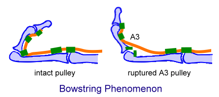
This results in inefficient flexion of the finger, reducing its range of motion.