• Skip to main content
  • Skip to primary sidebar
  • Skip to footer
  • General Ortho
  • Procedures
  • Spine
  • Upper Limb
  • Lower Limb
  • Pain
  • Trauma
  • Tumors
  • Newsletter/Updates
  • About Us
  • Contact Us

Bone and Spine

Orthopedic health, conditions and treatment

Ganglion Impar Block

By Dr Arun Pal Singh

In this article
    • Relevant Anatomy
    • Indications for Ganglion Impar Block
    • Ganglion Impar Block
    • Ganglion Impar Block with Modified Needle-Inside-Needle Technique
      • Risks and Complications

The ganglion impar is a  sacrococcygeal ganglion, also called ganglion of Wather. It is a retroperitoneal structure located at the level of the sacrococcygeal junction.

It is the final terminus of sympathetic chains formed by the fusion of both the chains.

Therefore, the ganglion impar block is used in patients with the pain in pelvic and perineal structures.

Relevant Anatomy

Sympathetic chain and ganglion impar

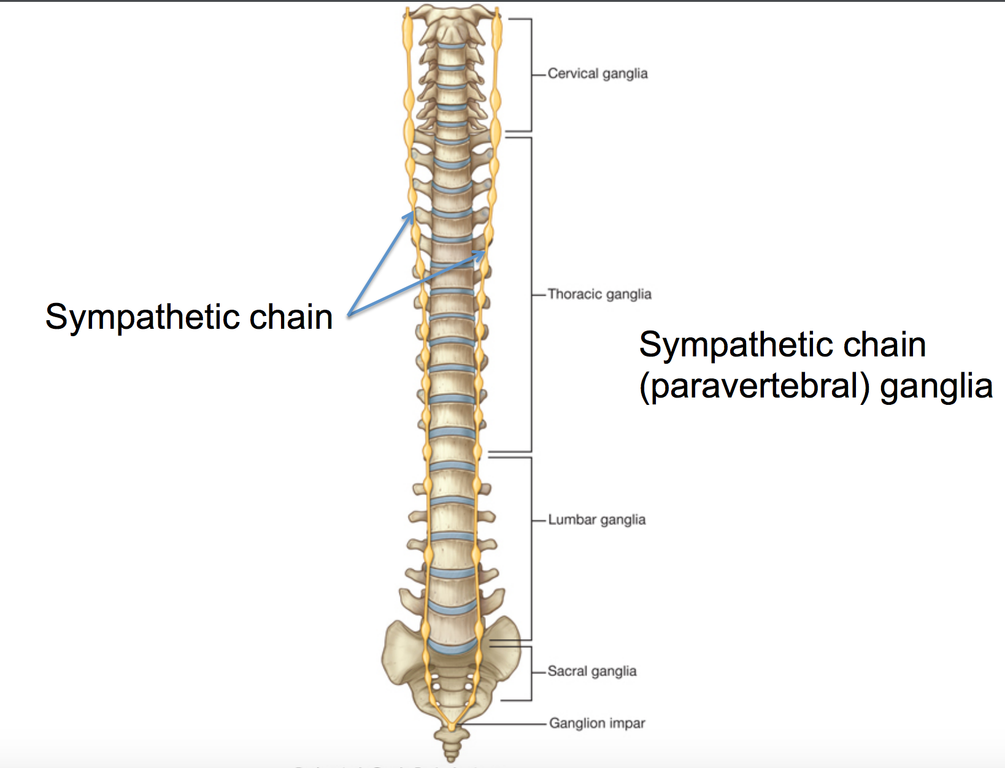
The ganglion impar is an irregularly shaped terminal ganglion of the sympathetic chain. It is located almost in the midline at the level of sacrococcygeal junction. It is of variable shape and can be located anywhere from the anterior surface of the sacrococcygeal junction to the lower coccygeal vertebral bodies.

The rectum is situated anterior to the ganglion. Fibers from the ganglion travel to the sacral spinal nerves along the gray rami communicantes.

This ganglion marks the end of the two sympathetic chains.

Visceral afferents from the perineum, distal part of the rectum, anus, distal end of the urethra, vulva, and distal third of vagina converge at the ganglion impar.

There is a wide range of variation in location of the ganglion.

Indications for Ganglion Impar Block

In orthopedics, ganglion impar block is used for coccydynia not amenable too other measures.

Other indications for this block are

  • Intractable perineal pain
    • Rectal/Anal pain (proctitis)
    • Vulvar pain
    • Vaginal pain [distal one-third]
    • Postherpetic neuralgia
    • Pain  in complex regional pain syndrome
  • Visceral pain
    • Endometriosis
    • Chronic prostatitis

Ganglion Impar Block

The classical technique was described by Plancarte and colleagues. Here, the patient is placed in lateral decubitus position with hips fully flexed.

A standard 22-gauge, 3.5-inch spinal needle is bent 1 inch from its hub to form a 30-degree angle. The needle is then introduced under local anesthesia through the anococcygeal ligament with its concavity oriented posteriorly, and under fluoroscopic guidance, it is directed along the midline at or near the sacrococcygeal junction.

A finger is placed in the rectum to avoid puncturing the structure.

A curved needle was later used instead of a bent needle. Spinal needle has also been advocated.

Various approaches to ganglion impar
Various approaches to ganglion impar, SCJ – Sacrococcygeal joint, ICJ- Intracoccygeal joint

Technical difficulties and the procedure associated risks [needle break, rectal perforation] have made this approach unpopular.

Transcoccygeal approach is more commonly used now. This involves the placement a 20-gauge, 1.5-inch needle through the sacrococcygeal ligament under fluoroscopic guidance so that the tip of the needle is just anterior to the anterior portion of the sacrum, thus minimizing the risk of rectal perforation.

The transsacrococcygeal ligament needle-inside-needle is the most frequently used approach.

A needle-in-needle technique may avoid trauma to the disk and coccyx, in addition to decreasing needle breakage during the transcoccygeal approach

Imaging guidance markedly increased technical success rates. Fluoroscopy facilitated further development, including the transcoccygeal, transarticular, transdiscal, intracoccygeal, and paracoccygeal approaches.

Ultrasound-guided techniques provide the advantage of real-time visualization and avoidance of radiation.

There are four main anatomic approaches to consider when performing ganglion impar blocks:

Paracoccygeal approaches i.e., immediately to the right or left of the coccyx have also been described.

Ganglion Impar Block with Modified Needle-Inside-Needle Technique

  • The patient is kept in a prone position
  • Keep a pillow under the abdomen
    • Allow flexion of the lumbosacral spine
    • Allows internal rotation of lower limbs
  • Prepare the skin of the area
  • Use a local anesthetic to numb the skin over the insertion point using a 25-gauge needle over the sacrococcygeal disc
  • Insert a 21-gauge, 1.5-inch needle through the sacrococcygeal disc under fluoroscopic guidance
    • Serves as guide for the other needle
  • Introduce a 25-gauge, 2-inch through the 22-gauge needle.
  • Confirm the needle placement in the retroperitoneal space by contrast agent insertion [0.2-0.5]. The comma sign confirms the placement.
  • For diagnostic block/prognostic block
    • 1 to 3 mL of local anesthetic (lignocaine, ropivacaine or bupivacaine) is injected.
  • For neurolysis
    • 1- 2 mL of a neurolytic agent [ 99% alcohol, phenol]
    • Given 20 to 30 minutes after the diagnostic block.

It is important that if pa atient complains pain with the neurolytic agent, the injection is stopped immediately.

Absolute bed-rest is indicated after neurolytic injection without position change. This is done to avoid the an unwanted spread of neurolytics to surrounding structures.

Risks and Complications

o             Rectal perforation and tract contamination

o             Infection

o             Fistula

o             Needle break


Spread the Knowledge
  • 6
    Shares
  •  
    6
    Shares
  • 6
  •  
  •  
  •  
  •  

Filed Under: Procedures

About Dr Arun Pal Singh

Arun Pal Singh is an orthopedic and trauma surgeon, founder and chief editor of this website. He works in Kanwar Bone and Spine Clinic, Dasuya, Hoshiarpur, Punjab.

This website is an effort to educate and support people and medical personnel on orthopedic issues and musculoskeletal health.

You can follow him on Facebook, Linkedin and Twitter

Primary Sidebar

drug resistant tuberculosis

Drug Resistant Tuberculosis – Types and Treatment

A person is said to have drug-resistant tuberculosis if the tuberculous bacteria that the person is infected with, will not respond to, and are resistant to, at least one of the main antitubercular drugs. Drug-resistant tuberculosis occurs when bacteria become resistant to the drugs used to treat TB. This means that the drug can no […]

muckle wells syndrome

Muckle Wells Syndrome

Muckle-Wells syndrome [Also known by the names of  UDA (Urticaria Deafness Amyloidosis) syndrome   is a rare autosomal dominant disease which results in Sensorineural deafness Recurrent hives Amyloidosis – A disease where abnormal deposition of amyloid occurs in various organs. Fevers Painful joints (arthritis) Muckle-Wells syndrome was first described in 1962 by Thomas James Muckle and […]

Tubercular Arthrits of Hip

Musculoskeletal Tuberculosis – Symptoms and Treatment

Musculoskeletal tuberculosis (Tuberculosis of bone and joints or popularly known as bone TB) is found more in children, adolescents, and young adults, than in the elderly. The spine is the most common site of involvement, followed by the hip and knee. In the spine, the usual involvement is vertebral bodies and intervertebral disks in the […]

Lumbar Degenerative Disc Disease

Lumbar Degenerative Disc Disease

Lumbar degenerative disc disease refers to a condition in which a compromised or degenerative disc in lumbar spine causes low back pain. Lumbar degenerative disc disease is quite common. At least 30% of people aged 30-50 years old are estimated to have some degree of disc space degeneration. All patients with the degenerated disc are […]

Broken implant in spine injury, for TLICS

TLICS- Thoracolumbar Injury Classification and Severity Score

Thoracolumbar injury classification and severity score or TLICS  is a scoring system for thoracolumbar fractures which classifies and provides guidance to the treatment as well. TLICS is based on three major categories Morphology of the injury Integrity of the posterior ligamentous complex Neurologic status of the patient. Denis classification of spinal injuries which is based […]

osgood schlatter disease

Osgood Schlatter Disease Causes Symptoms and Treatment

Osgood Schlatter disease is osteochondrosis of tibial tubercle that results from repetitive quadriceps contraction through the patellar tendon at its insertion on tibial tubercle. This occurs in the period before puberty as the tibial tubercle is growing and is susceptible to strain. It is is a self-limiting disorder of the knee. It is named after […]

Bertolotti syndrome is caused by lumbosacral transitional vertebra.

Bertolotti Syndrome – Causes and Treatment

Bertolotti syndrome is the presence of a variation of the fifth lumbar vertebra with a large transverse process, either articulated or fused with the sacral basis or iliac crest, producing a chronic, persistent lower back pain. It may also cause isolated L4/5 disc disease. Bertolotti described a unilateral and bilateral enlargement of the transverse process […]

Browse Articles

Footer

Pages

  • About
    • Policies
    • Contact Us

Featured Article

Shoulder Joint Anatomy

The shoulder is actually composed of four joints, namely glenohumeral joint, acromioclavicular joint, sternoclavicular joint and scapulothoracic … [Read More...] about Shoulder Joint Anatomy

Search Articles

© Copyright: BoneAndSpine.com