Last Updated on October 29, 2023
The gluteal region is an anatomical area located posteriorly to the pelvic girdle, at the proximal end of the femur. In approximate terms, it can be called the area of buttocks.
There are two gluteal regions, left and right.
The muscles in this region move the lower limb at the hip-joint.
Cutaneous Nerves, Vessels and Lymphatics
The cutaneous nerves
- The upper and anterior part is supplied by the lateral cutaneous branches of the subcostal and iliohypogastric nerves.
- The upper and posterior part is supplied by the posterior rami of spinal nerves L1,2,3 and S1,2,3.
- The lower and anterior part is supplied by branches from the posterior division of the lateral cutaneous nerve of the thigh.
- The lower and posterior part is supplied by branches from the posterior cutaneous nerve of the thigh (S1,2,3) and the perforating cutaneous nerve.
The blood supply of the skin and subcutaneous tissue is derived from perforating branches of the superior and inferior gluteal arteries.
The lymphatics from the gluteal region drain into the lateral group of the superficial inguinal lymph nodes.
Deep Fascia
Below the skin and subcutaneous tissue is deep fascia which is above and in front of the gluteus maximus (i.e. , over the gluteus medius) is thick, dense, opaque and pearly white. Over the gluteus maximus, however, it is thin and transparent. The deep fascia splits and encloses the gluteus maximus muscle.
Muscles of Gluteal Region
The muscles of the gluteal region can be broadly divided into two groups:
Superficial [Abductors and extenders]
A group of large muscles that abduct and extend the femur. Includes the gluteus maximus, gluteus medius, gluteus minimus , nd tensor fascia lata.
Deep [Lateral rotators]
A group of smaller muscles that mainly act to laterally rotate the femur. These include the quadratus femoris, piriformis, gemellus superior, gemellus inferior and obturator internus.
Superficial Muscles of Gluteal Region

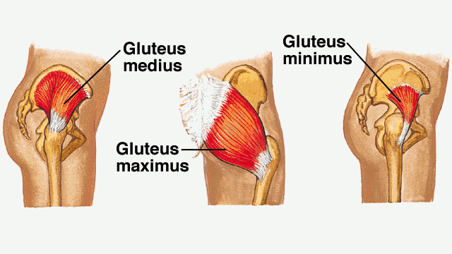
Gluteus Maximus
The gluteus maximus is the largest and the most superficial muscle. It produces the shape of the buttocks.
Origin
The muscle originates from
- Outer slope of the dorsal segament of iliac crest.
- Posterior gluteal line.
- Posterior part of gluteal surface of ilium behind the posterior gluteal line.
- Aponeurosis of erector spinae.
- Dorsal surface of lower part of sacrum.
- Side of coccyx.
- Sacrotuberous ligament.
- Fascia covering gluteus maximus.
Insertion
It slopes across the buttock at a 45 degree angle, then inserts into the iliotibial tract and the gluteal tuberosity of the femur. [ The deep fibers of the lower part of the muscle are inserted into the gluteal tuberosity. And the greater part of the muscle is inserted into the iliotibial tract.]
Nerve Supply
Inferior gluteal nerve.
Action
Gluteus maximus is the major extensor of hip joint and also assists with lateral rotation. It is used in forceful activities like running or climbing.
Gluteus Medius
The gluteus medius is fan-shaped and lies underneath the gluteus maximus and covers the lateral surface of the pelvis and hip. It lies between gluteus maximus and the minimus.
Origin
Gluteal surface of ilium between the anterior and posterior gluteal lines.
Insertion
Into the greater trochanter of femur, on oblique ridge on the lateral surface. The ridge runs downwards and forwards.
Nerve Supply
Superior gluteal nerve (L5,S1)
Actions
Gluteus medius abducts and medially rotates the lower limb. During locomotion, it secures the pelvis, preventing pelvic drop of the opposite limb.
Posterior fibres of the gluteus medius are also thought to produce a small amount of lateral rotation.
Nerve Supply
Superior gluteal nerve.
Gluteus Minimus
The gluteus minimus is the deepest and smallest of the superficial gluteal muscles. It is covered by the gluteus medius.
It is similar is shape and function to the gluteus medius.
Origin
From gluteal surface of ilium between the anterior and inferior gluteal lines.
It inserts into the greater trochanter of femur, on a ridge on the lateral part of the anterior surface.
Nerve Supply
Superior gluteal nerve (L5,S1)
Actions
Abducts and medially rotates the lower limb. During locomotion, it secures the pelvis, preventing pelvic drop of the opposite limb.
Tensor Fascia Lata
Tensor fasciae lata is a small superficial muscle which lies towards the anterior edge of the iliac crest. It lies between the gluteal region and the front of the thigh.
Origin
- Anterior 5 cm of the outer lip of the iliac crest up to the tubercle.
- Anterior superior iliac spine, and the notch below it.
Insertion
Iliotibial tract iliotibial tract, which itself attaches to the lateral condyle of the tibia.
Nerve Supply
Superior gluteal nerve (L4,5)
Actions
It assists the gluteus medius and minimus in abduction and medial rotation of the lower limb.
The Deep Muscles of Gluteal Region

Deep gluteal muscles is a group of smaller muscles lying underneath the gluteus minimus. They are lateral rotators of hip and stabilise the hip joint by pulling’ the femoral head into the acetabulum of the pelvis.
Piriformis
The piriformis is the most superior of the deep muscles. It lies, below and parallel to the posterior border of the gluteus medius.
Origin
It arises within the pelvis from.
- Pelvis surface of the middle three pieces of the sacrum, by 3 digitations.
- Upper margin of the greater sciatic notch and the adjoining areas of the sacroiliac joint and of the sacrotuberous ligament.
Insertion
The rounded tendon is inserted into the apex of the greater trochanter of the femur.
Nerve Supply
Nerve to piriformis [Ventral rami of S1,2].
Actions
Lateral rotation and abduction.
Obturator Internus
The obturator internus forms the lateral walls of the pelvic cavity. Some authors consider the obturator internus and the gemelli muscles as one muscle – the triceps coxae.
Origin
It is a fan-shaped, flattened belly lies in pelvis and the tendon in the gluteal region which arises from
- Pelvis surface of obturator membrane.
- Pelvis surface of the body of the ischium, ischial tuberosity, ischiopubic rami, and ilium below the pelvic brim.
Insertion
The tendon of the obturator internus leaves the pelvis through the lesser sciatic foramen and runs laterally to be inserted into the medial surface of the greater trochanter of the femur.
Nerve Supply
Nerve to obturator internus (L5,S1)
Actions
Lateral rotation and abduction.
Superior and Inferior Gemelli
The gemelli are two narrow and triangular muscles. They are separated by the obturator internus tendon.
Origin
The superior gemellus muscle originates from the ischial spine, the inferior from the ischial tuberosity.
Insertion
Both the gemelli are inserted on the greater trochanter of femur
Nerve Supply
Superior gemellus – Nerve to obturator internus
Inferior gemellus – Nerve to quadratus femoris
Actions
Lateral rotation and abduction.
Quadratus Femoris
The quadratus femoris is most inferior of the deep gluteal located below the gemelli and obturator internus.
Origin
It is flat, square-shaped muscle. Which lies between inferior gemellus and adductor magnus. It arises from
- Upper part of the outer border of ischial tuberosity.
Insertion
Quadrate tubercle and the area below it on the intertrochanteri crest.
Nerve Supply
Nerve to quadrates femoris (L5,S1)
Actions
Lateral rotation.
Obturator externus
Obturator externus is a triangular muscle that covers the outer surface of the anterior wall of the pelvis.
Origin
It arises from
- Outer surface of obturator membrane.
- Outer surface of the bony margins obturator foramen
Insertion
The muscle ends in a tendon which runs upwards and laterally behind the neck of the femur to insert into the trochanteric fossa on medial side of the greater trochanter.
Nerve Supply
Posterior division of obturator nerve. [L3,4]
Structures Under Gluteus Maximus
Muscles
- Gluteus medius
- Gluteus minimus
- Reflected head of the rectus femori
- Piriformis
- Obturator internus with two gemelli
- Quadratus femoris
- Obturator externus
- Origin of the four hamstrings from the ischial tuberosity
- Insertion of the upper or pubic fibres of the adductor magnus.
Vessels
- Superior gluteal vessels
- Inferior gluteal vessels
- Internal pudendal vessels
- Ascending branch of the medial circumflex femoral artery
- Trochanteric anastomosis
- Cruciate anastomosis
- First perforating artery
Nerves
- Superior gluteal nerve
- Inferior gluteal nerve
- Sciatic Nerve
- Posterior cutaneous nerve of the thigh
- Nerve to the quadrates femoris
- Pudendal nerve
- Nerve to the obturator internus
- The perforating cutaneous nerves
Bones and joints
- Ilium
- Ischium with ischial tuberosity
- Upper end of femur with the greater trochanter
- Sacrum and coccyx
- Hip joint
- Sacroiliac joint.
Ligaments
- Sacrotuberous
- Sacrospinous
- Ischiofemoral
Bursae
- Trochanteric bursa
- Ischial Bursa
- Bursa between the gluteus maximus and vastus lateralis.
Structures Deep to the Gluteus Medius
- Superior gluteal nerve
- Deep branch of the superior gluteal artery
- Gluteus minimus
- Trochanteric bursa
Structures Lying Deep to the Gluteus minimus
- Reflected head of the rectus femoris
- Capsule of the hip joint.
Vessels of the Gluteal Region
Superior Gluteal Artery
Superior gluteal artery is a branch of the posterior division of the internal iliac artery. It enters the gluteal region through the greater sciatic foramen. It passes above the piriformis along with the superior gluteal nerve. In the foramen, it divides into superficial branch that supplies the gluteus maximus.
The deep branch subdivides into superior and inferior branches, which run along the anterior and inferior gluteal lines respectively, between the gluteus medius and the gluteus minimus. The superior branch ends at the anterior superior iliac spine by anastomosing with the ascending branch of the lateral circumflex femoral artery and with branches from the deep circumflex iliac artery.
The inferior branch joins the trochanteric anastomosis [Discussed below]
Inferior Gluteal Artery
Inferior gluteal artery is branch of the anterior division of the internal iliac artery. It enters the gluteal region by passing through greater sciatic foramen, below the piriformis [Note that superior gluteal artery is above the piriformis], along with the inferior gluteal nerve.
It supplies
- Muscular branches to the gluteus maximus and to all muscles deep to it below the piriformis
- Cutaneous branches to the buttock and the back of the thigh
- Articular branch to the hip joint
- Branch to cruciate anastomotic branch
- Artery to the sciatic nerve
- Coccygeal branch which supplies the area over the coccyx.
Internal Pudendal Artery
It is also a branch of the anterior division of the internal iliac artery. It enters the gluteal region through the greater sciatic foramen and has a very short course in the gluteal region.
It crosses the ischial spine just medial to the nerve to the obturator internus, and lateral to the pudendal nerve. It leaves the gluteal region by passing into the lesser sciatic foramen to reach ischiorectal fossa.
Anastmosis in Gluteal Region
Trochanteric Anastomosis
Trochanteric anastomosis is present near the trochanteric fossa. It supplies branches to the head of the femur.
It is formed by
- Inferior division of the deep branch of the superior gluteal artery
- Ascending branch of the medial circumflex artery
- Ascending branch of the lateral circumflex artery
- Inferior gluteal artery.
This anastomosis is a channel of communication between the internal iliac and femoral arteries.
Cruciate Anastomosis
This anastomosis is situated over the upper part of the back of the femur at the level of the middle of the lesser trochanter.
It is formed
- Anastomotic branch of the inferior gluteal artery
- Ascending branch of the first perforating artery
- Transverse branch of the medial circumflex femoral artery
- Transverse branch of the lateral circumflex femoral artery.
This anastomosis is a connection between the internal iliac and femoral arteries.
Nerves of Gluteal Region
Spinal segments that the nerve carries are indicated in parenthesis.
Superior Gluteal Nerve (L4,5,S1)
Superior gluteal nerve is a branch of the sacral plexus which enters the gluteal region above the piriforms through the greater sciatic foramen, runs forwards between the gluteus medius and minimus.
It supplies the gluteus minimus and the tensor fasciae latae.
Inferior gluteal Nerve (L5,S1,2)
This is also a branch of the sacral plexus which enters the gluteal region through the greater sciatic foramen below the piriformis. It supplies the gluteus maximus.
Sciatic nerve (L4,L5,S1,S2,S3)
Sciatic nerve is the main continuation of the sacral plexus and thickest nerve in the body. It enters the gluteal region through the greater sciatic foramen, below the piriformis.
It runs downwards between the greater trochanter and the ischial tuberosity and enters the back of the thigh at the lower border of the gluteus maximus where it continues its course further in the lower limb.
Posterior Cutaneous Nerve of the Thigh (S1, S2, S3)
It is a branch of the sacral plexus. It enters the gluteal region through the greater sciatic foramen, below the piriformis and runs downwards medial or posterior to the sciatic nerve. It continues in the back of the thigh immediately deep to the deep fascia.
Branches
- Perineal branch which supplies the skin of the posterior two-thirds of the scrotum, or labium majus
- Gluteal supply the skin of the posteroinferior quadrant of the gluteal region.
Nerve to Quadratus Femoris (L4,L5, S1)
This nerve arises from the sacral plexus, enters the gluteal region through the greater sciatic foramen, below the piriformis, and runs downwards deep to the sciatic nerve, the obturator internus, and the gemelli.
It supplies the quadratus femoris, the gemellus inferior and the hip joint.
Pudendal Nerve
A branch of the sacral plexus that enters the gluteal region through the greater sciatic foramen and crosses the ischial spine to enter the ischiorectal fossa.
It supplies the obturator internus and the gemellus superior.
Nerve to the Obturator Internus
This is a branch of the sacral plexus and follows a similar course as pudendal nerve. It supplies the obturator internus and the gemellus superior.
Perforating Cutaneous Nerve (S2,S3)
It is a branch of the sacral plexus which pierces the lower part of the sacrotuberous ligament, winds around the lower border of the gluteus maximus, and supplies the skin of the posteroinferior quadrant of the gluteal region.
Sciatic Foramina and Structures Passing Through Them

Sacrospinous and sacrotuberous ligaments convert the greater and lesser sciatic notches of the hip bone into greater and lesser foramina respectively.
Following structures pass through these foramina

Greater Sciatic Foramen
The piriformis emerging from the pelvis fills the foramen almost completely. Structures are mentioned as passing above the piriformis or below it.
Structures passing above the piriformis
- Superior gluteal nerve
- Superior gluteal vessels
Structures passing below the piriformis are
- Inferior gluteal nerve
- Inferior gluteal vessels
- Sciatic nerve
- Posterior cutaneous nerve of thigh
- Nerve to quadratus femoris
- Pudendal nerve
- Internal pudendal vessels
- Nerve to obturator internus.
Lesser Sciatic Foramen
- The upper and lower parts of the foramen are filled up by origins of the two gemelli muscles.
- Tendon of obturator internus
- Pudendal nerve
- Internal pudendal vessels
- Nerve to obturator internus.
Clinical Significance of Gluteal Region
Intramuscular Injections

Intramuscular injections are given in the anterosuperior quadrant of the gluteal region, ie., in the glutei medius and minimus.
Weakness of Gluteus Maximus
Paralysis of gluteal muscle weakens the extension of the hip. The patient is not able to stand up from a sitting posture without support. Such patients, while trying to stand up, rise gradually, supporting their hands first on the legs and then on the thighs. This climbing on oneself is a frequently seen feature in muscular dystrophies.
Weakness of Gluteus medius and minimus
These muscles are principal hip abductors and important in stabilizing pelvis when a person walks.
If weak, the affected person has to sway on the paralyzed side to clear the opposite foot off the ground to compensate for lack of function. The person walks with lurching gait. Bilateral affection leads to waddling gait.
Normally when the body weight is supported on one limb, the glutei of supported side raise the opposite (and unsupported) side of the pelvis. However, if the abductor mechanism is defective, the unsupported side of the pelvis drops [Positive Trendelenburg’s test]. The extra sway towards the affected side is used to compensate the drop and improve foot clearance.