• Skip to main content
  • Skip to primary sidebar
  • Skip to footer
  • General Ortho
  • Procedures
  • Spine
  • Upper Limb
  • Lower Limb
  • Pain
  • Trauma
  • Tumors
  • Newsletter/Updates
  • About Us
  • Contact Us

Bone and Spine

Orthopedic health, conditions and treatment

Stress Views of Xrays

By Dr Arun Pal Singh

In this article
    • How To Obtain Stress Views?

Stress views are special x-ray views in which radiograph of the part examined is done after applying requisite stress. Stress views are important in the evaluation of ligamentous tears, joint stability, and fracture unions.

For example, stress views in the hand are usually ordered if there is an injury to the ulnar collateral ligament of the first metacarpophalangeal joint [Gamekeeper’s thumb].

Similarly, in the lower extremity,  the stress views of the knee and ankle joints are frequently obtained.

How To Obtain Stress Views?

Stress views are done after applying the force and direction of the force depends on the part examined and the purpose of the x-ray.Varus Stress Views knee

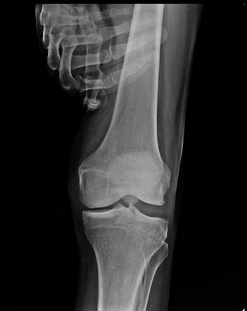
For example, if one wants to evaluate the integrity of the lateral collateral ligament [Lateral collateral ligament connects femur bone to the tibia across the knee joint, on the lateral or outer aspect], the patient is asked to lie on his back and knees are flexed about 15° to 20°.

With the thigh fixed with one hand assistant applies pressure on the medial aspect of the leg so as to push the foot towards the other foot producing varus at the knee.

The joint space would open as shown in the image above.

The films thus obtained show the anteroposterior projection.

Evaluation of ankle ligaments also requires stress radiography.

A trained assistant who understands the forces and knows to maintain them while the x-ray is being shot is always required for stress views. With little education and training, this can be achieved easily.

One of the other situations where stress views are important is the evaluation of fracture union. Sometimes, the status of fracture union is not clear from routine xray projections.

In such cases, the forces are applied in opposite directions on either side of the fracture so as to cause a displacement. In the view taken, opening or closing of fracture is noted.

If such phenomenon is present, the fracture has not united as yet.

Image Credit

Radiopedia

Spread the Knowledge
  •  
  •  
  •  
  •  
  •  
  •  

Filed Under: Imaging

About Dr Arun Pal Singh

Arun Pal Singh is an orthopedic and trauma surgeon, founder and chief editor of this website. He works in Kanwar Bone and Spine Clinic, Dasuya, Hoshiarpur, Punjab.

This website is an effort to educate and support people and medical personnel on orthopedic issues and musculoskeletal health.

You can follow him on Facebook, Linkedin and Twitter

Reader Interactions

Leave a Reply Cancel reply

Your email address will not be published. Required fields are marked *

This site uses Akismet to reduce spam. Learn how your comment data is processed.

Primary Sidebar

Blood supply of spinal cord

Blood Supply of Spinal Cord

The blood supply of spinal cord is mainly dependent on three vessels. These are namely anterior median longitudinal arterial trunk and a pair of posterolateral trunks. These posterolateral trunks are situated near posterior nerve rootlets. These longitudinal arterial trunks are largest in size in the cervical and lumbar regions because the demand for grey matter […]

pulled elbow

Pulled Elbow or Subluxation of Radial Head in Children

Pulled elbow is the term for subluxation of the radial head that affects children younger than 6 years, typically occurring due to a quick pull on a child’s arm. It is also called nursemaid elbow or babysitter’s elbow. It is an easily treatable condition. The left arm is more commonly involved, presumably because most caretakers […]

wrist pain text image

Wrist Pain Causes and Treatment

Wrist pain is a common problem and can be caused by many conditions, trivial and serious both. The pain can originate from carpal or wrist bones, ligaments, muscles, tendons, nerves, vessels or connective tissue and other structures in the vicinity. Damage to any of the parts of your wrist can cause pain and affect your […]

Fraser Classification of Floating Knee Injuries

Floating knee Injuries in Adults and Children

Floating knee refers to the fracture of ipsilateral [same side] fractures of tibia and femur. This makes knee unattached to tibia or femur, therefore name floating knee is given. The word floating knee was introduced for the first time by McBryde in 1965. Floating knee injuries may include a combination of diaphyseal, metaphyseal, and intra-articular […]

Blood supply of Talus

Blood Supply of Talus and Its Significance

Blood supply of talus was described in detail first by Wildenauer in 1950.   Since then many researchers have contributed. Talus is one of the few bones where the outcome of injury is affected due to injury to a unique pattern of the blood supply. Avascular necrosis of the talus is common after the injuries […]

repetitive strain injury

Repetitive Strain Injury – Causes, Symptoms and Treatment

Repetitive strain injury or RSI is a term describing the conditions where symptoms develop due to build of damage over the period, owing to repetitive motion. To say some person has repetitive strain injury or repetitive stress injury, other pathologies should not be present and the pain complaints can be related to repeated activities over […]

Discography in Lateral position Image

Lumbar Discography – Indications, Procedure and Inference

Lumbar discography is an invasive diagnostic procedure that involves, instillation of iodinated contrast into the nucleus pulposus after inserting a needle under fluoroscopy. This procedure is done with an aim to get information on the morphology and integrity nucleus pulposus, vertebral endplates and annulus fibrosus. In addition to this, the measured contrast injection allows determination […]

Browse Articles

Footer

Pages

  • About
    • Policies
    • Contact Us

Featured Article

PHILOS or Proximal Humerus Internal Locking System

PHILOS stand for Proximal Humerus internal locking system. It is an anatomic fixation system for the proximal humerus which also provides angular … [Read More...] about PHILOS or Proximal Humerus Internal Locking System

Search Articles

© Copyright: BoneAndSpine.com