Last Updated on August 2, 2019
Ulnar club hand is a congenital deformity of upper limb characterized by deficiency of the ulna and/or the ulnar sided carpal structures. The deformity can lead to instability of wrist or elbow.
Ulnar club hand is more appropriately termed as ulnar deficiencies of forearm. It is 5-10 times less common than radial club hand and is not associated with systemic conditions like radial club hand
About 70% of cases of ulnar club hand are unilateral.
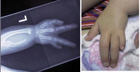
Image Credit: Orthobullets
Pathology of Ulnar Club Hand
Usually, the absence of ulna is partial rather than complete. The child has ulnar shortening with radial bowing. Digital anomalies can be found in about 90%.
The fourth and fifth metacarpals, as well as the capitate, lunate, triquetrum, hamate, and pisiform, are absent or deformed.
Ulnohumeral synostosis or radiohumeral synostosis can also be present. The radial head dislocation is present in about 50% of cases.
Radial bowing is produced by the tethering effect of the fibrocartilaginous ulnar anlage, which can also tether the carpus, producing limitation of wrist movement.
In most cases, however, the function is very good, and the carpus can be actively centralized.
Because thumb is present, the hand is more functional than radial club hand.
Most of the cases of ulnar club hand have sporadic occurrence although genetic syndromes are also known to be associated. Focal dermal hyperplasia, or Goltz [X-linked inheritance] , Split-hand and split-foot with ulnar dysplasia [autosomal dominant] and ulnar defect with mammary gland aplasia syndrome are to name a few.
Clinical Presentation of Ulnar Club Hand
There is a limb-length discrepancy of the upper limb and the function is affected. The condition is not painful.
On examination, there is a shortened, bowed forearm with loss of ulnar digits. Elbow function is affected.
Following findings may be noted and may vary from cases to case.
- Ulnar deviation of the hand
- Absent ulnar digits
- Syndactyly
- Elbow stability
- Elbow stiffness
- Limited pronation, supination, or both
- Radial head subluxation or dislocation
- Deficient carpal bones
- Stable wrist
All affected patients should undergo a detailed evaluation by a hand and upper limb therapist to assess hand function.
Classification of Ulnar Club Hand
Type 1
Ulnar shortening (distally) with a minor radial bow
- Hypoplasia of the ulna
- Proximal and distal epiphyses present
- Hypoplastic or absent ulnar digits
- Minimal radial bowing
Type 2
Significant ulnar shortening with a fibrocartilaginous anlage attached to the ulnar carpus, with significant radial bowing
- Partial aplasia of the ulna, distal third
- Distal ulnar anlage
- Bowed radius with anlage acting as a tether
- Presence/absence of progressive radial head dislocation
Type 3
Complete absence of the ulna
- Unstable elbow
- Straight radius
Type 4
The complete absence of the ulna, with a fibrocartilaginous anlage attached to the ulnar carpus
- Radiohumeral synostosis at the elbow
- Bowed radius
Imaging
X-rays
X-rays of the upper limb are done to evaluate elbow, forearm, wrist, and hand. Serial radiographs may be helpful in assessing the degree and course of the aplasia.
Magnetic Resonance Imaging
MRI can be used to evaluate the fibrocartilaginous anlage
Treatment of Ulnar Club Hand
Approach to patient
Initial management of ulnar club hand in infants consist of corrective casting and splinting. A long arm cast is applied with the limb in the corrected position. Cast needs to be changed periodically. Later on, removable splints can be used to maintain correction.
This treatment should be considered until the child is 6 months old. At this time exploration and excision of ulnar anlage should be done.
Surgical intervention is needed for syndactyly, radial bowing [ulnar anlage], dislocation of the radial head with limited elbow extension and forearm pronation and supination, and internal rotation deformity of the humerus.
Various treatment options are
For Ulnar deviation
Casting from birth, long-arm cast, gradual stretching of the tight ulnar structures. (Mild cases will correct by age 6 mo.)
Anlage
Anlage excision is done
Bowing of Radius
Radial corrective osteotomy. It is also useful to correct malrotation of the forearm
Radial Head Dislocation
- Observation only
- Resection only
- Resection with the creation of a single-bone forearm.
Unstable Elbow
Ulnohumeral or radiohumeral arthrodesis or elbow ligamentous reconstruction
Radiohumeral Synostosis
In young children excision of anlage suffices but in distal humeral osteotomy may also be needed.
Surgery is contraindicated in the presence of no or minimal functional deficit or any surgical risk factor due to associated syndromes or severely affected limb with poor neurologic function
Most of the children do well. Known complications are tightening of the flexor tendons to the digits causing restriction of hand function.
Dislocation of an unstable elbow is also a potential complication.