• Skip to primary navigation
  • Skip to main content
  • Skip to primary sidebar
bone and spine logo

Bone and Spine

Your Trusted Resource for Orthopedic Health Information

  • Home
  • About
  • Contact Us
  • Policies
  • Show Search
Hide Search
You are here: Home / Radiology and Imaging / Radiographs in Orthopedics

Radiographs in Orthopedics

Dr Arun Pal Singh ·

Last Updated on April 30, 2025

Radiographs are also called roentgenographs named after the discoverer of X-rays, Wilhelm Röntgen. Radiographs are produced by transmitting X-rays through a patient.  Rontgen discovered X-rays in 1895 and received the first Nobel Prize in Physics for their discovery in 1901. The x-ray is a very common term used for radiographs.

Contents hide
1 How is a Radiograph Done?
2 Different Views of Radiogrphas
3 Radiographs in Injuries
4 References

How is a Radiograph Done?

In the conventional film-screen radiography, an X-ray machine tube generates a beam of X-rays, aimed at the part of the patient to be radiographed.

The X-rays that pass through the patient are filtered through a device called an X-ray filter, to reduce scatter and noise, and strike an undeveloped film, which is held tightly to a screen of light-emitting phosphors in a light-tight cassette.

After the exposure, the film is developed chemically and an image appears on the film.

Film-screen radiography is being replaced by digital radiography, in which the X-rays strike a plate of sensors that convert the signals generated into digital information, which is transmitted and converted into an image displayed on a computer screen.

Radiographs of normal wrist

Radiography is often the first-line test of choice in radiological diagnosis and has a very important role in orthopedic diagnoses.

Different Views of Radiogrphas

It is important to obtain the right kind of x-ray to get to know further about a problem. Different parts of the body require different angles at which x-rays should be taken from.

These are called views.

It is essential to obtain correctly done views that show the areas of interest adequately.

Standard Views

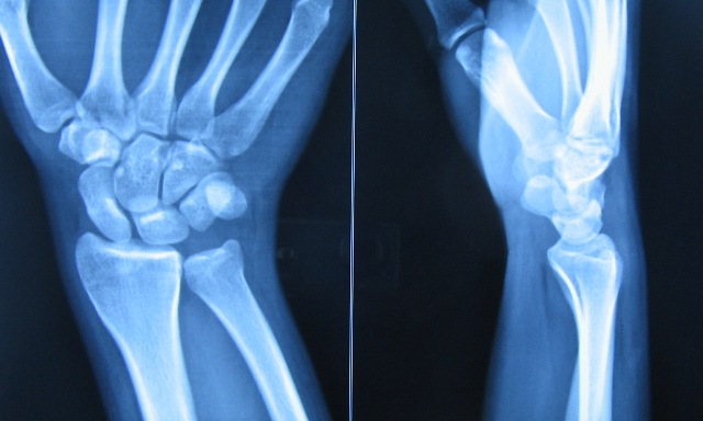
Routine standard views are anteroposterior and lateral views. In some body parts, one or more oblique films may be required. Roughly translated, front to back direction is anteroposterior or AP view, whereas sidewise view is lateral. Oblique views of radiographs lie somewhat in between.

Generally, films of long bones should show both proximal and distal joints. Thus if radiographs of the tibia are to be done, it should include both knee joint and ankle joint.

There are special views for just about any area and unconventional views can be improvised when required.

History and clinical examination of the patient tell the physician about part of the patient to be radiographed.

Comparative Views

Because the body is bilaterally symmetrical, we can have a radiograph of other/contralateral normal part for comparison. Comparison views are most needed in some pediatric cases because of variable growth rates and variable appearance of growth plates.

Stress Views

Stress views of radiographs are done after applying the stress in the desired direction. Stress view radiographs are done to assess joint stability or to assess the soundness of the union of the fracture.

Stress views are special x-ray views in which a radiograph of the part examined is done after applying requisite stress. Stress views are important in the evaluation of

  • ligamentous tears
  • joint stability,
  • fracture unions.

For example, stress views in the hand are usually ordered if there is an injury to the ulnar collateral ligament of the first metacarpophalangeal joint [Gamekeeper’s thumb].

For example, if one wants to evaluate the integrity of the lateral collateral ligament of knee, the patient is asked to lie on his back, and his knees are flexed about 15° to 20°.

With the thigh fixed with one hand assistant applies pressure on the medial aspect of the leg to push the foot towards the other foot producing varus at the knee.

The joint space would open up on the radiograph.

Evaluation of ankle ligaments also requires stress radiography.

Radiographs in Injuries

Radiographs remain the first investigation that is performed when an injury occurs.

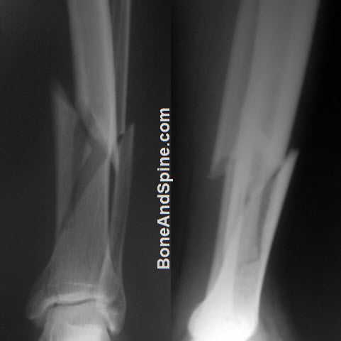
Typically a fracture would appear as a break in the continuity of the bone. But appearance may vary.

In adults, a cortical break is usually a complete one. It is visible like a crack in the intact bone or bone might appear divided into two parts.

Radiographs showing Comminuted Fracture of Lower End Tibia With Fracture of Fibula
Comminuted Fracture of Lower End Tibia With Fracture of Fibula

In contrast, in children there may be a partial cortical break as in greenstick fracture, buckling of cortex on one side as in torus fracture, or bending of the bone without a visible break on radiographs, called plastic fracture.

Impacted fracture is a type of fracture where it may show only an increased area of bone density, but usually, there is some disruption of the cortex.

In addition to the break in the bone,  there might be associated signs in the soft tissue. For example, there might be a break in the skin visible.

Above all, clinical correlation is most important. In case one finds that the radiographs do not correlate with clinical examination, one should proceed with better investigations.

References

  • Sy JW, Lopez AJ, Lausé GE, Deal JB, Lustik MB, Ryan PM. Correlation of stress radiographs to injuries associated with lateral ankle instability. World J Orthop. 2021 Sep 18;12(9):710-719. [link]
  • Andrew G. Geeslin, Diego F. Lemos, Matthew G. Geeslin. Knee Ligament Imaging: Preoperative and Postoperative Evaluation. Clinics in Sports Medicine. 2021,Volume 40, Issue 4, 657-675 [DOI]

Radiology and Imaging This article has been medically reviewed by Dr. Arun Pal Singh, MBBS, MS (Orthopedics)

About Dr Arun Pal Singh

Dr. Arun Pal Singh is a practicing orthopedic surgeon with over 20 years of clinical experience in orthopedic surgery, specializing in trauma care, fracture management, and spine disorders.

BoneAndSpine.com is dedicated to providing structured, detailed, and clinically grounded orthopedic knowledge for medical students, healthcare professionals, patients and serious learners.
All the content is well researched, written by medical expert and regularly updated.

Read more....

Primary Sidebar

Know Your Author

Dr. Arun Pal Singh is an orthopedic surgeon with over 20 years of experience in trauma and spine care. He founded Bone & Spine to simplify medical knowledge for patients and professionals alike. Read More…

Explore Articles

Anatomy Anatomy Fractures Fractures Diseases Diseases Spine Disorders Spine Disorders Patient Guides Patient Guides Procedures Procedures
featured image for orthopedics traction

Orthopedic Traction – Principles, Types, and Uses

Traction is a fundamental concept in orthopedics for managing …

featured image of gower sign for segmenatal instability of lumbar spine

Clinical Tests for Lumbar Segmental Instability

Lumbar segmental instability may not always be visible on standard …

mesurement of scoliosis for braces

Braces for Scoliosis- Types, Uses and Results

Braces for scoliosis are recommended to prevent the scoliotic curve …

discogenic back pain

Discogenic Back Pain Causes, Diagnosis and Treatment

Discogenic back pain is a common cause of axial low back pain [the …

Elbow arthrodesis using internal fixation

Elbow Arthrodesis- Indications, Methods and Complications

Elbow arthrodesis refers to the surgical fusion of the elbow joint. It …

Popular articles

Foot Sprain

Sprain Causes and Treatment

A sprain is an injury that occurs to …

Seinsheimer classification of subtrochanteric fractures

Subtrochanteric Fractures Femur- Presentation and Treatment

Subtrochanteric fractures are fractures …

triphalangeal thumb photo

Triphalangeal Thumb Presentation, Types and Treatment

The triphalangeal thumb is a congenital …

Meniscofemoral ligaments

 Meniscofemoral ligaments – Humphery and Wisberg Ligaments

Meniscofemoral ligaments are straight …

Bone and Spine

© 2025 BoneAndSpine.com · All Rights Reserved
The content provided on BoneAndSpine.com is intended for informational and educational purposes only. It is not a substitute for professional medical advice, diagnosis, or treatment. Read Disclaimer in detail.