• Skip to primary navigation
  • Skip to main content
  • Skip to primary sidebar
  • Home
  • Online Consultation
  • About
  • Newsletter/Updates
  • Contact Us
  • Policies

Bone and Spine

Orthopedic health, conditions and treatment

  • General Ortho
  • Procedures
  • Spine
  • Upper Limb
  • Lower Limb
  • Pain
  • Trauma
  • Tumors

Typical and Atypical Thoracic Vertebra

By Dr Arun Pal Singh

In this article
    • Typical Thoracic Vertebra
      • Attachments on a Typical Thoracic Spine Vertebra
    • Atypical Thoracic Vertebrae
      • First Thoracic Vertebra
      • Ninth Thoracic Vertebra
      • Tenth Thoracic Vertebra
      • Eleventh Thoracic Vertebrae
      • Twelfth Thoracic Vertebra
      • Related

The typical thoracic vertebrae are seven in number and atypical thoracic vertebra are five in number.

Vertebra T2 to T8 are typical and rest of them are atypical.

Both typical and atypical thoracic vertebrae are discussed.

Typical Thoracic Vertebra

Thoracic Vertebra

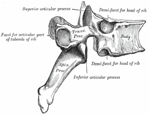
The body of a typical thoracic vertebrae is heart shaped with roughly the same measurements from side to side and anteroposteriorly. On each side it bears 2 costal facets (demifacets). The superior costal facets are larger and placed on the upper border of the body near the pedicle. They articulate with the head of the numerically corresponding rib. The inferior costal facets are smaller and placed on the lower border in front of the inferior vertebral notch. They articulate with the next lower rib.

The vertebral foramen is comparatively small and circular.

The vertebral Arch

  • The pedicles are directed straight backwards. The superior vertebral notch is shallow, while the inferior vertebral notch is deep and conspicuous.
  • The laminae overlap each other from above.
  • The superior articular processes project upwards from the junction of the pedicles and laminae. The articular facets are flat and are directed backwards and a little laterally and upwards. This direction permits rotatory movements of the spine.
  • The inferior articular processes are fused to the laminae. Their articular facets are directed forwards and slightly downwards and medially.
  • The transverse processes are large, and are directed laterally and backwards from the junction of the pedicles and laminae. The anterior surface of each process bears a facet near its tip, for articulation with the tubercile of the corresponding rib. In the upper 6 vertebrae, the costal facets on the transverse processes are concave and face forwards and laterally. In others the facets are flat and face upwards, laterally and slightly forwards.
  • The spine is long, and is directed downwards and backwards. The 5th to 9th spines are the longest, more vertical and overlap each other. The upper and lower spines are less oblique in direction.

Attachments on a Typical Thoracic Spine Vertebra

  • The upper and lower borders of the body give attachment, in front and behind respectively, to the anterior and posterior longitudinal ligaments.
  • The upper borders and lower parts of the anterior surfaces of the laminae provide attachment to the ligamenta flava.
  • The transverse process gives attachment to (i) the lateral costotransverse ligament (at the tip); (ii) the superior costotransverse ligament (lower border); the inferior costotransverse ligament (anterior surface); (iv) the intertransverse muscles (to upper and lower borders); (v) the levator costae (posterior surface).
  • The spines give attachment to the supraspinous and interspinous ligaments. They also give attachment to several muscles including the trapezius, the rhomboids, the latissimus dorsi, the serrate posterior superior and inferior, and many deep muscles of the back.

Atypical Thoracic Vertebrae

First Thoracic Vertebra

  • The body resembles that of a cervical vertebra. It is broad and not heart shaped. Its upper surface is lipped laterally and beveled anteriorly. The superior costal facet on the body is complete. It articulates with the head of the first rib. The inferior costal facet is a “demifacet” for the second rib.
  • The spine is thick, long and nearly horizontal.
  • The superior vertebral notches are well marked, as in cervical vertebrae.

Ninth Thoracic Vertebra

It resembles a typical thoracic vertebra except that the body has only the superior costal facets (demifacets). The inferior costal facets are absent.

Tenth Thoracic Vertebra

It resembles a typical thoracic vertebra except that the body has a single complete superior costal facet on each side, extending on to the root of the pedicle.

Eleventh Thoracic Vertebrae

  • The body has a single large costal facet on each side, extending on to the upper part of the pedicle.
  • The transverse process is small and has no articular facet. Sometimes it is difficult to differentiate between vertebrae T10 and T11.

Twelfth Thoracic Vertebra

  • The shape of the body, pedicles, transverse processes and spine are similar to those of a lumbar vertebra. However, the body bears a single costal facet on each side, which lies more on the lower part of the pedicle than on the body.
  • The transverse process is small and has no facet, but has superior, inferior and lateral tubercles.
  • The inferior articular facets are lumbar in type and are directed laterally (everted), but the superior articular facets are thoracic in type.

Related

Spread the Knowledge
11
Shares
 
11
Shares
110   

Filed Under: Anatomy

About Dr Arun Pal Singh

Arun Pal Singh is an orthopedic and trauma surgeon, founder and chief editor of this website. He works in Kanwar Bone and Spine Clinic, Dasuya, Hoshiarpur, Punjab.

This website is an effort to educate and support people and medical personnel on orthopedic issues and musculoskeletal health.

You can follow him on Facebook, Linkedin and Twitter

Primary Sidebar

Browse Articles

Classification of Acetabular Fractures

Acetabular Fractures Presentation and Treatment

Acetabular fractures are uncommon but important because they involve a major weight-bearing joint in the lower extremity.  Being intraarticular fractures, these fractures require anatomical reduction for good long-term function of the hip joint. A failure to achieve anatomical reduction could lead to the breakdown of the cartilage surface due to articular incongruity present leading to degenerative […]

Radial Club Hand

Radial Clubhand Diagnosis and Treatment

Radial clubhand is a term for a longitudinal deficiency of the radius and other structures on the radial side of forearm and hand. For example, absent thumb is a common accompaniment. Proximal deficiencies also can occur throughout the arm and shoulder girdle Radial club hand is found in 1 per 100000 people. Radial club hand […]

periosteal chondroma of femur

Chondroma – Presentation, Types, Diagnosis and Treatment

The term chondroma refers to a benign tumor of cartilage origin. The tumor contains cartilage cells that are similar to normal cartilage cells but contains vessels that differentiate it from normal cartilage. Chondromas may arise inside or outside the bone. These tumors are called enchondromas when they occur in the medullary canal of the bone. The […]

microfracture chondroplasty

Microfracture Chondroplasty

The microfracture chondroplasty is a technique to treat full-thickness cartilage defects that lead to the formation of new joint surface cartilage. It includes the debridement of the damaged area and puncturing the underlying bone with a micro-instrument or pick to allow bleeding from the bone. The resulting blood clot that fills the cartilage defect has […]

Ankle Arthrodesis

Ankle Arthrodesis

Ankle arthrodesis is performed more commonly than that of the hip or knee. It is also called ankle fusion. Most common indication for performing this surgery is arthritis following an injury to ankle also called post-traumatic arthritis. Other indications are Rheumatoid arthritis Infection Neuromuscular disorders Failed total ankle arthroplasty. Bone tumors around the ankle Before […]

rib-cage

Anatomy of Thoracic Cage or Rib Cage

The thoracic cage or rib cage is the skeletal framework that encloses the contents of the thorax. The thoracic cage or rib cage along with its contents form the thorax portion of the body. Roughly speaking, this is the area of the chest. The Thoracic cage consists of the 12 pairs of ribs with their […]

Synovial fluid analysis

Synovial Fluid Analysis And Interpretation

Synovial fluid analysis is often done to aid in the diagnosis of joint inflammation, pain, swelling, and/or fluid accumulation. It is withdrawn from the joint by a procedure called arthrocentesis. Synovial fluid is a thick, stringy fluid found in the cavities of synovial joints. It reduces friction between the articular cartilage and other tissues in […]

© Copyright: BoneAndSpine.com
Manage Cookie Consent
The site uses cookies. Please accept cookies for a better visiting experience.
Functional Always active
The technical storage or access is strictly necessary for the legitimate purpose of enabling the use of a specific service explicitly requested by the subscriber or user, or for the sole purpose of carrying out the transmission of a communication over an electronic communications network.
Preferences
The technical storage or access is necessary for the legitimate purpose of storing preferences that are not requested by the subscriber or user.
Statistics
The technical storage or access that is used exclusively for statistical purposes. The technical storage or access that is used exclusively for anonymous statistical purposes. Without a subpoena, voluntary compliance on the part of your Internet Service Provider, or additional records from a third party, information stored or retrieved for this purpose alone cannot usually be used to identify you.
Marketing
The technical storage or access is required to create user profiles to send advertising, or to track the user on a website or across several websites for similar marketing purposes.
Manage options Manage services Manage vendors Read more about these purposes
View preferences
{title} {title} {title}
 

Loading Comments...