Last Updated on April 24, 2022
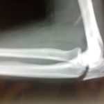
Olecranon fracture of olecranon process fracture is a fracture of the large curved eminence called the olecranon process that forms the point of the elbow. It is the point where we rest our elbow on the table in a sitting position.
Olecranon is a part of the ulna bone and is the uppermost posterior aspect of the ulna. Olecranon fractures range from simple nondisplaced fractures to complex fracture-dislocations of the elbow joint. Olecranon fracture is a common fracture due to the subcutaneous position of olecranon on the point of the elbow.
The olecranon process lies beneath the skin and it is very vulnerable to direct trauma.
The injury is less common in children.
The fracture has bimodal age distribution. It is caused by high energy injuries in the young and low energy injuries such as fall in the elderly
Relevant Anatomy of Elbow and Olecranon Process
 Together with the coronoid process, the olecranon forms the articular surface for articulation with the trochlea of the humerus. This articulation allows movement only in the anteroposterior plane.
The elbow is a hinge joint formed by distal humerus, proximal radius, and proximal ulna.
The olecranon is uppermost posterior part of the ulna that contributes to the articular surface of ulna [trochlear groove or notch], anterior contribution being by the coronoid process.
Medially, the trochlear part of the distal humerus articulates with the trochlear notch of the ulna formed by the olecranon process and coronoid process. Trochlear notch is a concavely shaped structure which participates in trochleohumeral joint and is responsible for flexion-extension movements of the elbow.
In the lateral part, the radius articulates with the humerus.
The elbow is held together by ligaments, muscles, and shape of the participating bones.
The elbow is a complex hinge joint. The major stabilizers to valgus are the medial (ulnar) collateral ligament and the radial head. The major stabilizer to varus stress is the lateral collateral ligament complex. The coronoid process stabilizes the humerus against the distal ulna.

Olecranon prevents anterior translation of the ulna with respect to the distal humerus. The anterior surface of the olecranon is covered with articular cartilage. Therefore, most of the fractures except the rare tip fractures are intra-articular fractures.
The triceps muscle inserts into the posterior third of the olecranon and proximal ulna. The periosteum of the olecranon blends with the triceps. Therefore, in olecranon fractures extension mechanism is affected.
Fracture displacement is largely due to the pull of the triceps. Usually, wide separation of fragments indicates an extensive tearing of the fibrous sheath in which the unopposed triceps is contracted, drawing the separated fragment upward.
Classification of Olecranon Fractures
Colton’s classification
Nondisplaced/Stable
- Displaced less than 2 mm,
- Exhibit no change in position with gentle flexion to 90 degrees or with extension against gravity.
Displaced fractures
A. Avulsion fractures
B. Transverse/oblique fractures
C. Isolated comminuted fractures
D. Fracture/dislocations
AO Classification
- Type A – Extra-articular fractures
- Type B – Intra-articular fractures
- Type C – Intra-articular fractures of both the radial head and the olecranon
Schatzker Classification
- Type A – Simple transverse fracture
- Type B – Transverse impacted fracture
- Type C – Oblique fracture
- Type D – Comminuted fracture
- Type E – More distal fracture, which actually is extra-articular
- Type F – Fracture dislocation
Mayo Clinic Classification
- Type I – Nondisplaced
- Type II – Displaced but stable
- Type III – Associated instability of the elbow
Presentation of Olecranon Fracture
The most common mechanism of an olecranon fracture is a fall on the semiflexed supinated forearm. Tense triceps snaps the olecranon over the lower end of the humerus, which acts as a fulcrum. A fall on the elbow or direct blow to the elbow could also result in an olecranon fracture.
Hyperextension injuries of the elbow, and forceful throws are less common causes of olecranon fractures.
Stress fractures have also been reported.
The patient presents with swelling of the posterior part of elbow. There is pain on the extension of the limb. Sometimes, a gap may be palpated when examining the continuity of the olecranon process.
Gross disability can be noted in displaced fractures.
On examination, the elbow is swollen and there is tenderness over the olecranon area. The mobility of the fragment may be noted. The patient would be unable to extend the elbow or it is painful in case of undisplaced fractures.
Elbow and the limb should be examined for associated injuries. Olecranon injury may be part of polytrauma and the patient should be assessed for other injuries in other regions as well.
The Extensor mechanism is almost always affected in displaced fractures and the patient is unable to extend the elbow.
Imaging
Olecranon fracture is best visualized in lateral view x-ray of the elbow. The following things are to be noted
- Fracture location and if reaches the articular surface
- Displacement and the gap
- Presence of comminution and degree of comminution
On the lateral view, the olecranon fractures appear as a lucency usually reaching the trochlear groove articular surface.
Following associated findings should be looked for presence or absence
- Fracture of the coronoid process
- Fracture or dislocation of the radial head
- Presence of fracture of the distal humerus
Treatment of Olecranon Fractures
The treatment of olecranon fractures varies with individuals.
In young persons, the goal is
- To restore articular surface and stability
- Preserving the motor power
- Prevention of joint stiffness.
In older patients, apart from the severity of the injury, the treatment is guided by the levels of activity.
In patients who have concomitant medical conditions, conservative treatment should be considered.
Indications for surgery in olecranon fracture are
- >2 mm of displacement
- Affected extensor mechanism
- Associated elbow instability
- Failure of non-operative treatment
Nonoperative Treatment
This treatment is carried in nondisplaced and stable olecranon fracture
These fractures are treated by immobilization in an above-elbow cast. Traditionally, the elbow was positioned in 30 degrees of flexion for fear of displacement of the fracture fragments. But long arm cast with the elbow in 90 degrees of flexion for 3 to 4 weeks works well too and is more comfortable for the patients.
The position of the fragments is checked after 5-7 days by x-ray of the elbow.
The cast is kept for 3-4 weeks and after that protected range of motion, exercises are begun and continued for another 4 weeks.
In the case of an elderly patient, the range of motion may be initiated earlier.
Operative Treatment
Open reduction and internal fixation is the treatment of choice displaced olecranon fractures. The choice of implant depends on the fracture pattern.
- Transverse fractures
- Oblique fractures
- intramedullary screw with or without tension band wiring over the screw.
- Plate fixation with a lag screw
- Severely comminuted fractures
- Plate fixation
- Excision and triceps advancement
- Severe Osteoporosis
- Excision and triceps advancement
After a stable fixation, the patient should be put on the range of motion exercises for the elbow after about 10 days.
In avulsion fractures of olecranon, a transverse fracture line separates a small proximal fragment of the olecranon process from the rest of the ulna. This fracture is most common in elderly patients and is usually caused by an avulsion force
If the fragment is small, excision is the best treatment with repair of the triceps back to the bone. If the fragment is large then tension band wiring surgery should be done.
Complications of Olecranon Fractures
- Hardware irritation
- Heterotopic ossification
- Non-union
- Infection
References
- Newman SD, Mauffrey C, Krikler S. Olecranon fractures. Injury. 2009 Jun. 40 (6):575-81. [link]
- Sultan S, Khan AZ. Management of comminuted fractures of the olecranon by tension band wiring. J Ayub Med Coll Abbottabad. 2003 Jul-Sep. 15 (3):27-9.
- Matar HE, Ali AA, Buckley S, Garlick NI, Atkinson HD. Surgical interventions for treating fractures of the olecranon in adults. Cochrane Database Syst Rev. 2014 Nov 26. 11:CD010144.

