Last Updated on March 15, 2020
The ulna is a long bone in the forearm. It lies medially and parallels to the radius, the other bone in the forearm
It acts as stabilizing the bone, with the radius pivoting to produce the pronation and supination movement of the forearm.
The bone participates in the formation of the elbow joint by articulating with the humerus. Distally, it articulates with the radius, forming the distal radio-ulnar joint.
It is the medial bone of the forearm and is homologous with the fibula of the lower limb. Along with other bone called radius, it connects elbow with the wrist.
The ulna is stabilizing bone of the forearm on which the radius can pronate and supinate for efficient working of the upper limb.
Structure of Ulna
Ulna has upper and lower ends and a shaft.
The Upper End
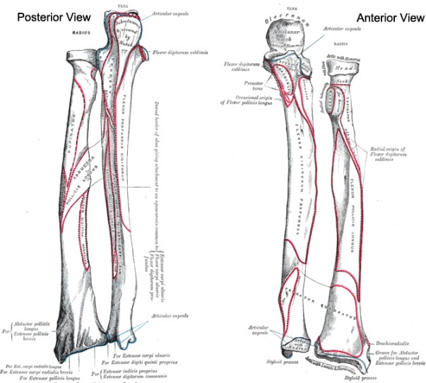
The upper end of ulna presents the olecranon and coronoid processes, and the trochlear and radial notches.

image credit: Teach Me Anatomy
Olecranon Process
The olecranon process projects upwards from the shaft. It has superior, anterior, posterior, medial and lateral surfaces. The anterior surface is peculiar. It is concave and forms the upper part of the trochlear notch. The posterior surface forms a triangular subcutaneous area which is separated from the skin by a bursa.
Inferiorly, it is continuous with the posterior border of the shaft. The upper part forms the point of the elbow. The medial surface of the shaft. The lateral surface is continuous inferiorly with the posterior surface of the shaft.
Coronoid Process
Coronoid process projects forwards from the shaft just below the olecranon and has four surfaces, superior, anterior, medial and lateral. The superior surface forms the lower part of the trochlear notch. The anterior surface is triangular and rough. Its lower corner forms the ulnar tuberosity.
The upper part of its lateral surface is marked by the radial notch for the head of the radius.
The lower part of the lateral surface forms a depressed area (to accommodate the radial tuberosity). It is limited behind by a ridge called the supinator crest.
Trochlear Notch
It forms an articular surface that articulates with the trochlea of the humerus to form the elbow joint.
Radial Notch
The radial notch articulates with the head of the radius to form the superior radioulnar joint.
Shaft
The shaft has three borders and three surfaces.
Borders
The interosseus (or lateral) border is sharpest in its middle two fourths. Inferiorly it can be traced to the lateral side of the head. Superiorly it is continuous with the supinator crest.
The anterior border of the shaft is thick and rounded. It begins above on the medial side of the ulnar tuberosity, passes backward in its lower one third, and terminates at the medial side of the styloid process.
Surfaces
Ulna has three borders anterior, posterior and medial.
Distal Portion
The distal end is much smaller in diameter than the proximal end. It terminates in a rounded head which also has a distal projection – the ulnar styloid process.
The head articulates with the ulnar notch of the radius to form the distal radio-ulnar joint.
Attachments
Muscles
Triceps
The common tendon of triceps brachii muscle inserts on the posterior part of the superior surface of the olecranon process
Anconeus muscle
Anconeus muscle inserts on the lateral aspect of the olecranon process.
Brachialis Muscle
Brachialis muscle inserts on to the interior surface of the coronoid process.
Pronator Teres
Pronator teres muscle originates from the medial surface on the middle portion of the coronoid process. It shares the origin with medial epicondyle of the humerus.
Flexor carpi ulnaris muscle
This muscle takes origin from the olecranon process and posterior surface. It also shares the origin with medial epicondyle of the humerus.
Flexor Digitorum
Flexor digitorum superficialis and profundus muscles take origin from the coronoid process and the anteromedial surface of ulna
Pronator Quadratus
It originates from the distal portion of the anterior ulnar shaft.
Extensor carpi ulnaris
It originates from the posterior border of ulna [and lateral epicondyle of the humerus]
Supinator muscle
Originates from the proximal region [along with lateral epicondyle of the humerus]
Abductor pollicis longus
This muscle originates from the posterior surface [and also from the posterior surface of the radius bone]
Extensor pollicis longus
It originates from the dorsal shaft [and also from the dorsal shaft of the radius and the interosseous membrane)
Extensor Indicis
It originates from the posterior surface of the distal part (and interosseous membrane)
Other Attachments
Interosseous Membrane
It attaches to interosseous border.
Elbow Capsule
Capsular ligament of the elbow joint attaches to the margins of the trochlear notch i.e. coronoid process and olecranon process.
Annular Ligament
The annular ligament is attached to the anterior and posterior margins of the radial notch.
Ulnar Collateral Ligament
Ulnar collateral ligament of the wrist attaches to the styloid process.
Articular Disc
The articular disc of the inferior radioulnar joint is attached by its apex to a small rough area just lateral to the styloid process.
Clinical Significance
Injuries
Forearm bones fracture very commonly. Often both bones of forearm fracture but isolated fractures of ulna also occur.
The Olecranon process gets fractured when a patient falls on a flexed elbow. The triceps brachii can displace part of the fragment proximally.
Dislocation of the elbow occurs when there is a fall on an outstretched hand.
Monteggia’s fracture and Galeazzi’s fracture are other common injuries that involve ulna.
Madelung Disease
It is a deformation of the distal end of the radius and leads to the dorsal subluxation of the ulna.