Last Updated on October 27, 2023
Wound care dressings and procedures aim at rapid covering healing of skin defects are for better wound healing.
The best method for healing a wound is to close the defect by surgical means as quickly after the injury as possible. However, this may not always be possible.
Where primary closure is not possible [ large surface, very deep wounds], following things take priority
- Keep wound infection free
- Reduce or eliminate pain
- Remove or reduce all potential inhibitors of natural healing (eg, Dead tissue, Fibrosis etc)
- Replace or substitute the missing tissue as much as possible.
When the injury occurs, the aim of the treatment is to oppose the wound ends so as the wound heals by primary intention. When this is not possible, the wounds heal mostly by secondary intention.
In such cases appropriate wound dressings are necessary.
Classification of Wounds
Based on the Thickness of the Wound
- Superficial Wounds – Involve only the epidermis and the upper dermis
- Partial-thickness Wounds- Skin loss up to the lower dermis.
- Full-thickness Wounds- Involve the skin and the subcutaneous tissue.
- Deep Wounds – Deep wounds include complicated wounds with laceration of blood vessels and nerves, wounds penetrating into natural cavities, and wounds penetrating into an organ or tissue
Based on Involvement Structures
- Simple- Comprise only one tissue.
- Combined- Involve two or more tissues.
Based on Time Elapsed
- Fresh – Less than 8 hours from the trauma
- Old – More than 8 hrs
Based on Morphology
- Excoriation or scarification [It is the most superficial type]
- Incised wound [As in surgical incision]
- Crush wound [Generally occurs with a heavy blow]
- Contused wound
- Lacerated wound [Fragments of tissue are torn as result of sharp injury]
- Slicing wound [ As in detachment of epicranial epineurosis.]
- Stab wound [Made with a pointed tool or a weapon]
- Gunshot Wound
- Animal/human bite
Based on Healing
- Primary Healing- When edges are approximated. Minimal scarring .Most surgical wounds heal by primary intention healing.
- Delayed Primary Healing- This type of wound healing occurs when a wound is initially cleaned, debrided, and observed, typically 4 or 5 days before closure. Many wounds which are initially contaminated are purposely left open and a delayed closure is done.
- Healing By Secondary Intention In this type of healing, a full-thickness wound is allowed to close and heal. Secondary healing results in an inflammatory response that is more intense than with primary wound healing. A larger amount of granulation tissue is formed and there is a greater contraction. This kind of healing leads to larger scars.
- Healing by Epithelization Epithelization is a part of wound healing occurs in all kinds of wound healing as part of the phases of wound healing. It is the process by which epithelial cells migrate and replicate and traverse the wound. In wounds involving only the epidermis and superficial dermis, epithelization is the predominant method by which healing occurs.
Wound Healing
Wound healing is a complex process taken up by the body to repair the defect by restoring cellular structure and tissue layers. The wound healing process has distinct phases which are
- Hemostasis – The mechanisms to control bleeding kick in
- Inflammatory Phase- The immune cells and inflammatory markers are released at the wound site,
- Proliferative or Granulation Phase- Healing of the bone by making new scar tissue
- Remodeling Phase- The tissue is remodeled to match the original tissue characteristics
Wound Coverings
Efficient wound dressings are desirable for both small and large wounds. Currently, available wound dressings or coverings can be divided into two categories
- Permanent coverings, such as autografts
- Temporary coverings, such as allografts, xenografts, and synthetic dressings.
Autografts are considered best for wound covering on a healthy tissue but a major disadvantage of autograft is the creation of additional wound at donor side subjecting the patient to additional morbidity, the risk of infection and fluid/electrolyte imbalance, contour defects or scarring.
Temporary wound dressings do not increase patient morbidity but due to their synthetic or chemical components, limited persistence on the wound surface, and foreign body character.
Wound Bed Preparation
To achieve optimal healing, wounds should
- not be infected
- contain as much vascularized bed as possible
- be free of exudate.
To achieve this, the wound should be appropriately prepared to enhance both the effectiveness of the dressing and the self-healing ability of the wound.
Wound bed preparation is defined as the global management of the wound to accelerate endogenous healing or to facilitate the effectiveness of other therapeutic measures.
Acute and chronic wounds differ in preparation of their wound bed. For example, in acute wounds, debridement is effective for removal of damaged tissue and potential bacteria but for chronic wounds, where much more than debridement needs to be addressed for optimal results.
A well-vascularized wound bed is achieved by
- Removing necrotic or fibrous tissue
- Controlling edema
- Decreasing bacterial burden
- Compression therapy (especially for venous ulcers)
- Off-loading (in the setting of pressure-induced ulcers).
- Application of growth factors like platelet-derived growth factor [PDGF])
- Applying bioengineered skin
- Using occlusive dressings.
Autologous Skin Grafts
The best coverage is the patient’s own skin (ie, split-thickness skin grafts) from an adjacent matched undamaged area. However, the procedure inflicts an injury on the donor site.
Autograft is the method of choice to achieve definitive coverage of burned skin with good quality of the healed skin.
Allogenic Skin Grafts
The use of allogeneic cadaver skin as a biologic dressing is now widely accepted and is usually preferred to synthetic dressings. The preservation of allografts can be performed by different techniques, such as freeze-drying, glutaraldehyde fixation, or glycerolization.
Wound debridement or debridement is the process of removing infected, damaged, dead, nonviable tissue or foreign matter wounds so as to improve the healing potential of the remaining healthy tissue.
Wound debridement can be achieved with surgery or by nonsurgical means.
Types of Wound Dressings
The ideal wound dressing should be nontoxic, nonsensitizing, nonallergic, easy to use, provide mechanical and bacterial protection, maintain a moist environment at the wound/dressing interface, allow gas and fluid exchange, should not adhere to wound and cost-effective.
Dressings include
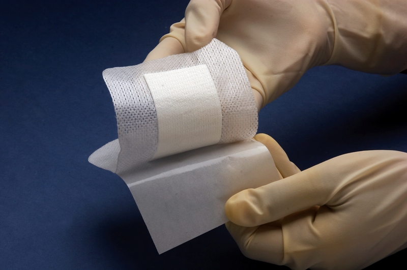
- Dry dressings- gauze and bandages, nonadhesive meshes, membranes and foils, foams, and tissue adhesives
- Moisture-keeping dressings – pastes, creams and ointments, nonpermeable or semipermeable membranes or foils, hydrocolloids, hydrogels, and combination products.
- Bioactive dressings include antimicrobial dressings, interactive dressings, and combination products.
- Skin substitutes include epidermal substitutes, acellular skin substitutes, autologous and allogenic skin, and skin substitutes containing living cells.
Different types of wounds require different dressings or combinations of dressings.
Skin substitutes are discussed below.
Acellular Dressings and Skin Substitutes
Amniotic membrane
Amniotic membranes can be applied on superficial second-degree burns, donor sites, and deep second-degree burns after early debridement. They have to be changed daily and need to be covered with gauze. They do not allow long-term coverage. Amniotic membranes can be kept refrigerated for 6 weeks, or they can be frozen for longer storage and banking purposes.
Acellular human dermis
Acellular human dermis substitute is essentially healthy human dermis with all the cellular material removed. It is then virus-screened and preserved by freeze-drying. The acellular human dermis is prepared from cadaver skin by extensive washing and purification followed by high-dose x-ray radiation.
Xenogenic Grafts [Grafts of animal origin]
Sterilized porcine skin is the most common source of the xenograft.
Acellular Matrices and Combination Products
Collagen, collagen containing products and collagen nylon combination sheets are used for covering the wound.
Synthetic or Semisynthetic dressings
Silicones
Silicone dressings are chemically and biologically inert, usually transparent. These are used as sheets or gels. Some of the silicone membranes are porous whereas other silicone membranes are nonpermeable. While former allow environmental exchange with the wound latter to ensure a fully occlusive wound environment.
Silicone wound dressings are antiadhesives and help to reduce hypertrophic and keloid scarring. Silicone has been found to be useful in flattening of scarring tissue; increasing elasticity; and reducing discoloration, making the scars more cosmetically acceptable.
Barrier Films
Barrier films are protective polymers dissolved in a fast-drying carrier solvent. These protect skin from losing moisture or from exogenic fluids, protect from skin stripping, and be compatible with clothing. They are available in liquid form which quickly polymerizes and membranes.
Foams
Foams are suitable for use on light-to-medium exuding wounds and not recommended for dry wounds. Anatomically shaped dressings are available for specific wound locations like sacral region or heel.
Tissue adhesives
Tissue adhesives are used in small and not-too-deep wounds that can heal by primary intention. These contain cyanoylate components which polymerize in an exothermic reaction on contact with either a fluid or a basic substance leading to the formation of a strong, flexible, waterproof band.
Tissue adhesives are used for simple lacerations.
Hyaluronic Acid
Hyaluronic acid films are indicated for use on diabetic foot ulcers and venous leg ulcers.
Hydrogels
Hydrogel dressings are suitable for use at all stages of wound healing except for infected or heavily exuding wounds and are a good alternative for classic wet dressings.
Hydrocolloids
Hydrocolloid dressings are constituted of a variety of constituents, such as methylcellulose, pectin, gelatin polyisobutylene. and some of them also contain alginate.
After contact with the wound surface, hydrocolloids slowly absorb fluids, leading to the formation of gel covering the wound. These help in keeping the wound moist, promote the granulation tissue and provide pain relief. Silicones are used in a dressing of acute wounds and chronic wounds, for desloughing, and exuding wounds. Depending on exudate levels, the dressing might be required to change frequently.
Calcium Alginates
Alginates are absorbable, biodegradable dressings containing mannuronic acid and glucuronic acid derived from seaweed. These are used in exuding wounds like leg ulcers, pressure sores, and infected surgical wounds.
They control exudate by ion exchange and also activate macrophages to generate proinflammatory signals like tumor necrosis factor-alpha, interleukin 1, interleukin 6 and interleukin 12. Alginates should not be applied to dry or drying wounds.
Dressings containing an antimicrobial agent
These dressings include the now sustained-release iodine and silver dressings
Local collagen-based drug delivery systems
Collagen-based dermal matrices (sponges or transparent, semipermeable membranes) containing gentamicin have been successfully used for the treatment of infected burn wounds and for the prevention of wound infection in large-surface wounds.
Engineered Skin Dressings and Substitutes
Allogenic Cultured Epidermal Sheets
Cultured allografts transplanted on deep partial-thickness skin burns induce faster healing of the wound.
Matrices Containing Living Skin-derived Cells
This is living cell containing bilayered product designed to speed up healing of both partial-thickness wounds and full-thickness wounds, including diabetic leg ulcers. It has recently been approved both in the United States and Europe.
Non-collagen–based products to Enhance Dermal Regeneration
Dermagraft is a Smith & Nephew product containing cryopreserved human fibroblast-derived dermal substitute composed of fibroblasts, extracellular matrix, and a bioabsorbable scaffold. Dermagraft is indicated for use in the treatment of full-thickness diabetic foot ulcers,
Hyaluronic acid products have been used alone or as a carrier for living cells. In this case, the patient’s own cells have been cultivated in vitro and seeded on the membrane prior to clinical application. Laserskin autograft is an epidermal substitute consisting of autologous keratinocytes on a laser-microperforated membrane of hyaluronic acid and is indicated for use on diabetic foot ulcers and venous leg ulcers.
BioSeed-S is an autologous skin graft for treating poorly healing wounds. For BioSeed-S treatment, a small piece of the patient’s own skin has to be removed. The skin cells are isolated and grown in the BioTissue laboratories. After about 2-3 weeks, the cells in fibrin adhesive (biological tissue glue) are applied to the patient’s wound.