Last Updated on April 11, 2022
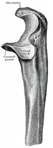
Coronoid fracture or coronoid process fracture is fracture anterior projection of ulna [ called coronoid] in its superior part where together with the posterior projection of ulna, olecranon, it expands the articular area of the upper end of the ulna. Ulna articulates with the lower end of humerus in this articular notch.
Both coronoid and olecranon processes also provide stability to humerus by forming anterior and posterior supports by virtue of projections.
While fracture of olecranon is very common, a coronoid fracture is seen less commonly. Fractures of the coronoid process usually reflect severe trauma to the elbow.
Coronoid fracture usually occurs in association with other injuries of the elbow, namely elbow dislocation. Isolated coronoid fracture is less common.
Large coronoid fractures often are associated with persistent elbow instability even after reduction of the dislocation.
Coronoid fracture accounts for less than 1-2% of all elbow fractures. Coronoid fractures have been identified in 10-15% of elbow dislocations.
Anatomy of Coronoid Process
Read more about Elbow anatomy

Coronoid Anatomy and Relevant Biomechanics
coronoid tip is an intraarticular structure. Along with the olecranon process, it acts as a humerus stabilizer.
The medial facet of the coronoid provides insertion for the medial ulnar collateral ligament and is important for varus stability
The Coronoid acts as an anterior buttress of the olecranon and is important in preventing recurrent posterior subluxation.
The Coronoid acts as the primary restraint of elbow subluxation or dislocation.
Mechanism of Coronoid Fracture
Previously, it was thought that these fractures were a result of avulsion force acting on the coronoid but now it is thought that they probably are due to the direct impact of trochlea on the coronoid when a force acts. Thus coronoid fractures result from shear injury.
Nothing inserts on coronoid process tip. Therefore avulsion theory is not sound.
The brachialis muscle, which was thought to put a force of avulsion, inserts much distally than the tip of the coronoid. However, it is not uncommon to find attached fibers of brachialis in a larger fragment distal to the coronoid process.
When the fragment of coronoid is large, it may cause dislocation of the elbow as the supportive buttress is no longer available [See classification]. But a small coronoid potentially severe trauma with possible acute recurrent dislocation.
Classification of Cornoid Fracture
The classification was suggested by Regan and Morrey who after a retrospective study, classified the fracture into three types.
 Classification of coronoid fracture
Type I—small avulsion fracture at tip of coronoid
Type II—fragment involves 50% of the coronoid but does not extend to the base
Type III—Fracture at the base of the coronoid, likely including the insertions of the brachialis and the anterior band of the medial collateral ligament.
In spite of their type, the presence of a coronoid fracture should evoke concern for acute instability.
Associated Injuries
Most coronoid fractures occur in associated with other injuries of the elbow. These associated injuries are
- lateral collateral ligament disruption [results from varus deforming force]
- Olecranon fracture-dislocation
- Radial head fractures and lateral collateral ligament injury [together]
- Terrible triad of elbow
- Coronoid fracture
- Radial head fracture
- Elbow dislocation
Imaging
AP and lateral views of the elbow are sufficient for most of the cases. In complex injuries of the elbow with concomitant coronoid fracture, CT may be required.
Treatment of Coronoid Fracture
Fractures of the coronoid process are generally not amenable to closed treatment. Therefore open reduction is almost always the treatment of choice where the fragment is stabilized using either screw or wire fixation.
Nonoperative treatment can be considered in fractures which have minimal displacemnt and elbow is stable.
However, because coronoid fractures are generally associated with elbow instability, open reduction with internal fixation of a significant coronoid fracture often provides the necessary stability to prevent further dislocation.
Operative intervention may also be considered for those fractures that interfere with joint motion. This can occur if the fragment is intraarticular or if it unites proximally and forms a significant bone block to flexion.
For the internal fixation, following options can be used
- Cerclage wire/screw for Type I injuries
- Cannulated screw fixation or plating for type II or III injuries ORIF
Lateral ligament repair for posteromedial rotatory instability should be done in type II or III.
The coronoid can be approached by medial or posterior approach. Posterior approach is used when there is fracture of olecranon too.
postoperative rehabilitation depends on intraoperative exam following the procedure thermoplastic resting splint applied with elbow at 90° and forearm in neutral restrict terminal 30° extension for 2-4 weeks avoid shoulder abduction for 4-6 weeks to prevent varus moment on arm early active motion
After the surgery, the limb is immobilized for a period of 3 to 4 weeks with elbow at 90 degrees of flexion and neutral rotation. Shoulder abduction should be avoided for 4-6 weeks as well.
Weekly follow-up should be done with x-ray.
After this period, gentle elbow mobilization is begun.
Complications of Coronoid Fracture
- Neurovascular injury
- Stiffness of elbow
- Heterotopic ossification
- Instability
- Recurrent dislocation
- Posttraumatic arthritis of the elbow
References
- Ring D, Horst TA. Coronoid Fractures. J Orthop Trauma. 2015 Oct. 29 (10):437-40. [ Link].
- Kovacevic D, Vogel LA, Levine WN. Complex Elbow Instability: Radial Head and Coronoid. Hand Clin. 2015 Nov. 31 (4):547-56.
- Ring D. Fractures of the coronoid process of the ulna. J Hand Surg Am. 2006 Dec. 31 (10):1679-89.
- Zhao S, Zeng C, Yuan S, Li R. Reconstruction of coronoid process of the ulna: a literature review. J Int Med Res. 2021 Apr. 49 (4):3000605211008323. [link]