Last Updated on October 27, 2023
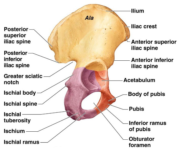
Hip bone is also known as innominate bone or pelvic bone and is formed by the fusion of three bones namely ilium, ischium and pubis bones. Hip bone forms part of the pelvis and takes part in hip joint articulation.
The term hip bone can create confusion in the mind of the reader. To understand better, it is important to differentiate between the hip and pelvis.
The pelvis is a ring-shaped structure formed by two innominate bones on front and side and the sacrum behind.
Hip is a joint formed between femur bone and acetabulum, a cavity in the innominate bone.
The innominate bone which is formed by the fusion of three bones has been called hip bone but actually, it is part of the pelvis [and contributes acetabulum for hip joint.]
The hip bone is made up of the three parts – the ilium, pubis, and ischium. Prior to puberty, a cartilage called the triradiate cartilage separates these individual bones. At the age of 15-17, the three parts begin to fuse.
Their fusion forms a cup-shaped socket known as the acetabulum, which becomes complete at 20-25 years of age. The head of the femur articulates with the acetabulum to form the hip joint.
Hip bones participate in the formation of pelvis along with sacrum as shown below in the diagragm.
[Read more about pelvis anatomy.]
Each hip bone, therefore, has three articulations –
- Sacroiliac joint – Articulation with the sacrum
- Pubic symphysis – Articulation with the other hip bone.
- Hip joint – Articulation with the head of the femur.
Side determination of Hip Bone
The side of the hip bone can be determined by keeping the following features in mind.
- The acetabulum is on the lateral side
- Obturator foramen lies below the acetabulum, pubis being anterior and ischium posterior
- Flat expanded part, the ilium is above the acetabulum.
Anatomical Position of Hip Bone
- Pubic tubercle and anterior superior iliac spine lie in the same coronal plane.
- Pelvic surface of pubis is directed backward and upwards
- The symphyseal surface of the body of pubis is in the median plane.
We now will discuss the individual bones that fuse to form the hip bone.
Ilium
Ilium is the largest part of the hip bone and forms expanded plate in the upper part and contributes to acetabulum formation in the lower part.
Roughly two-fifths of the acetabulum is contributed by ilium.
The upper end of the ilium is called iliac crest. The iliac crest is a broad, convex, topmost portion of ilium which can be palpated in the flank area. [Roughly where the trouser belt is the anterior
The anterior end of iliac crest is called the anterior superior iliac spine which is a very important anatomical landmark.
Iliac crest ends posteriorly in the posterior superior iliac spine [Located about dimple of venus or 4 cm lateral to the second sacral spine.]
Iliac crest is divided into a ventral segment and a dorsal segment which meet at the tubercle. The ventral segment forms more than the anterior two-thirds of the crest. The dorsal segment forms less than the posterior one-third of the crest.
The lower end is fused with ischium and pubis at the acetabulum.
Ilium has got three borders [anterior, posterior and medial] and three surfaces [gluteal, iliac, and a sacropelvic surface].
Borders of Ilium
Anterior border of ilium starts at the anterior superior iliac spine and runs downwards to the acetabulum. In the upper part, the border has a notch and while its lower part has the anterior inferior iliac spine.
Posterior border extends from the posterior superior iliac spine to the upper end of the posterior border of the ischium. It is marked by prominence called posterior inferior iliac spine and lower a large deep notch called the greater sciatic notch.
Medial Border is on the inner surface of the ilium from the iliac crest to the iliopubic eminence and separates the iliac fossa from the sacropelvic surface. Its lower rounder part forms the iliac part inlet of the pelvis or arcuate line.
Surfaces of Ilium
The gluteal surface is the outer surface of the ilium, which is convex in front and concave behind. Three gluteal lines divide the gluteal surface.
- The posterior gluteal line is the shortest and extends from a point front of the posterior superior spine to a point of the posterior inferior spine.
- The anterior gluteal line is the longest, begins about an inch behind the anterior superior spine, runs backwards and then downwards to end at the middle of the upper border of the greater sciatic notch.
- The inferior gluteal line is not well defined. It begins a little above and behind the anterior inferior spine, runs backwards and downwards to end near the apex of the greater sciatic notch.
The inner surface of the ilium is divided by medial border into iliac fossa and sacropelvic surface.
Iliac fossa is the large concave area on the inner surface of the ilium, situated in front of its medial border. It forms the lateral wall of the false pelvis.
Sacropelvic surface is the uneven area on the inner surface of the ilium, behind its medial border.
It is subdivided in three parts: the iliac tuberosity, auricular surface, and pelvic surface.
The iliac tuberosity is a large rough area below dorsal segment of the iliac crest. It is raised in the middle and depressed both above and below. Articular surface is called auricular surface and articulates with the corresponding auricular surface on sacrum to form sacroiliac joint.
Anteroinferior to the auricular surface is a smooth pelvic surface which forms lateral wall of the true pelvis. The preauricular surface is marked by a preauricular sulcus.
Attachments on Ilium


Anterior Superior Iliac Spine
ASIS provides attachment to the inguinal ligament. Sartorius muscle originates from ASIS and area below that.
Iliac Crest
Outer lip of iliac crest gives origin to fascia lata in whole extent and tensor fascia lata in front of the tubercle.
Latissimus dorsi muscle takes origin just behind the highest point of the crest. Anterior two-thirds of crest provides origin to external oblique muscles.
Inner lip of iliac crest gives rise to transverses abdominis muscle and fascia transversalis in anterior part and quadratus lumborum in posterior part.
The dorsal segment of iliac crest has following attachments
- Gluteus maximus arises from the lateral slope
- Erector spinae arises from the medial slope
- Interosseus and dorsal sacroiliac ligaments are attached to medial margin deep to erector spinae
- Anterior inferior iliac spine gives origin to the straight head of rectus femoris and iliofemoral ligament.
- Sacrotuberous ligament gives origin to upper fibers of sacrotuberous ligament and few fibers of the piriformis.
Gluteal surface
- Gluteus maximus is a big muscle and its upper fibers take origin from the area behind the posterior gluteal lines.
- From area between anterior and posterior gluteal lines, gluteus medius arises.
- Gluteus minimus arises from an area between anterior and inferior gluteal lines.
- Rectus femoris reflected head arises from groove above the acetabulum.
- The capsule of the hip joint is attached to acetabular margin.
Iliac Fossa
- Iliac fossa gives origin to iliacus in upper two thirds whereas lower one third is covered by the iliac bursa.
- Iliac tuberosity provides attachments to interosseus sacroiliac ligament, dorsal sacroiliac ligament and iliolumbar ligament superiorly.
- Margin of auricular surface gives attachment to ventral sacroiliac ligament.
Pelvic surface
Preauricular sulcus provides attachment to lower fibers of the ventral sacroiliac ligament, few fibers of piriformis and upper half of obturator internus.
Pubis
Pubis forms anteroinferior part of the hip bone contributes to anterior one-fifth of the acetabulum and forms the anterior boundary of the obturator foramen.
Pubis consists of body, superior ramus, and inferior ramus.
Body of pubis
It is flattened anteroposteriorly and has a border superiorly called pubic crest which ends in a pubic tubercle laterally. In males, the tubercle is crossed by the spermatic cord.
It has three surfaces
- The anterior surface is directed downwards, forward and slightly laterally. It is rough on the superomedial aspect.
Posterior surface or pelvic surface is directed upwards and backwards and forms the anterior wall of the true pelvis. - Medial surface or symphyseal surface articulates with opposite pubic symphysis.
Superior and inferior rami are extensions from the body of the pubis.
Superior Ramus
Three borders
- Superior border [also called pectin pubis or pectineal line] is sharp and extends from pubic tubercle to posterior aspect of iliopubic eminence. It forms part of the arcuate line [pelvic inlet].
- The anterior border is called the obturator crest. This border is a rounded ridge, extending from the pubic tubercle to the acetabular notch.
- The inferior border is sharp and forms the upper margin of the obturator foramen.
Three surfaces
- The pectineal surface is a triangular area between the anterior and superior borders, extending from the pubic tubercle to the iliopubic eminence.
- The pelvic surface lies between the superior and inferior borders. It is smooth and is continuous with the pelvic surface of the body of the pubis. The pelvic surface is crossed by ductus deferens in males and round ligament of uterus in females.
- The obturator surface or inner surface lies between the anterior and inferior borders. It presents the obturator groove. Obturator groove transmits obturator vessel and nerves.
Inferior Ramus
Inferior ramus extends from the body of the pubis to the ramus of the ischium, medial to the obturator foramen. It unites with the ramus of the ischium to form the conjoined ischiopubic rami. [It is discussed separately below after the ischium]
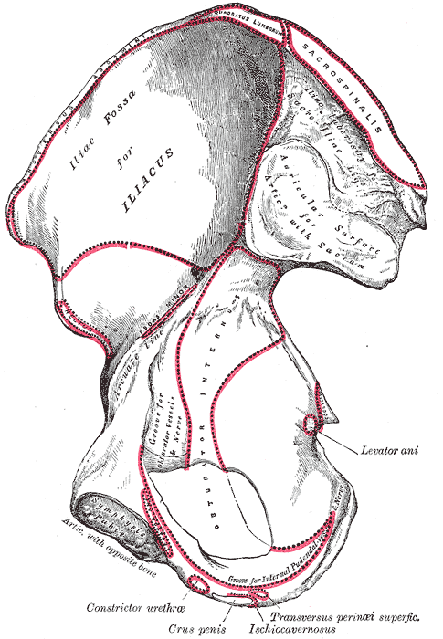
Attachments on the Pubis
Pubic Tubercle
Medial end of the inguinal ligament and ascending loops of the cremaster muscle.
Pubic Crest
The lateral part of the crest gives origin to the lateral head of the rectus abdominis , and to the pyramidalis. Medial head of the rectus abdominis which takes origin from pubic symphysis crosses the medial part of the pubic crest.
Body of Pubis
Anterior surface
- Anterior pubic ligament medially
- Adductor longus – From the angle between the crest and the symphysis
- Gracilis – From the margin of the symphysis, and from the inferior ramus
- Adductor brevis originates lateral to the gracilis
Posterior surface
- Levator ani from the middle part
- Obturator internus laterally
Superior Ramus
The pectineal line provides attachment to
- Conjoint tendon
- Medial end of the lacunar ligament
- Pectinate ligament
- Pectineus muscle and its covering fascia.
- Psoas minor [when present].
- Pectineal surface, in its upper part, gives rise to pectineus muscle.
Ischium
The ischium is posteroinferior part of the hip bone and forms two-fifths of the acetabulum and posterior body of the obturator foramen.
Ischium has a body and a ramus.
Body of Ischium
The body of the ischium is below and behind acetabulum and ends in ischial tuberosity. Its anterior border forms post margin of the obturator foramen.
Posterior border continues with post border of the ilium and takes part in the formation of the lower border of the greater sciatic notch and gives out a projection below that called ischial spine. Below that is the lesser sciatic notch. The lesser sciatic notch is occupied by the tendon of the obturator internus and a bursa deep to the tendon. The notch is lined by hyaline cartilage.
Dorsal surface of the body of the ischium is continuous with the gluteal surface of the ilium. Piriformis, the sciatic nerve, and the nerve to the quadratus femoris are closely related to this surface. The groove transmits the tendon of the obturator internus.
The surface between anterior and lateral borders is called femoral surface.
The pelvic surface is smooth and takes part in the lateral wall of the true pelvis [the lateral wall of the ischiorectal fossa.]
Ischial tuberosity has upper and lower areas demarcated by a transverse ridge. An oblique ridge further divides the upper area into a superolateral and inferomedial area. The inferomedial area is covered with fibrofatty tissue which supports body weight in the sitting position.
The lower area of ischial tuberosity is divided into inner and outer areas by a longitudinal ridge.
Ramus of Ischium
Ramus of ischium combines with inferior pubic ramus to form conjoined ischiopubic rami which is discussed below.
Conjoined Ischiopubic Rami
The inferior ramus of the pubis unites with the ramus of the ischium on the medial side of the obturator foramen. The site of the union may be marked by a localized thickening. The conjoined rami have two borders- upper and lower, and two surfaces – outer and inner.
The upper border forms part of the inferior margin of the obturator foramen and lower border forms the pubic arch along with the corresponding border of the bone of the opposite side.
The inner surface is convex and smooth. It is divided into upper middle and lower areas by two ridges.
Attachments on the Ischium
Ischial Spine
- Sacrospinous ligament along its margins
- The origin for the coccygeus and posterior fibers of levator ani from its pelvic surface.
Lesser Sciatic Notch
The upper and lower margins of the notch give origin to the superior and inferior gemelli.
Femoral Surface of the Ischium
- Origin to obturator externus along the margin of the obturator foramen
- Quadratus femoris along the lateral border of the upper part of the ischial tuberosity.
Ischial Tuberosity
- Superolateral part – origin to the semimembranosus
- Inferomedial area – Semitendinosus and the long head of the biceps femoris
- Inferolateral area – adductor magnus.
- Sacrotuberous ligament on medial margin
- Ischiofemoral ligament on the lateral border, just below the acetabulum.
Pelvic Surface
- obturator internus.
Conjoined ischiopubic rami
- Upper border – obturator membrane
- Lower border – Fascia latae, membranous layer of superficial fascia of the perineum, also known as Colle’s fascia.
- Outer surface
- Obturator externus [ near the obturator margin of both rami]
- the adductor brevis and gracilis [mainly from the pubic ramus]
- the adductor magnus [mainly from the ischial ramus]
- Inner surface
- Upper ridge – upper layer of the urogenital diaphragm.
- Lower ridge – perineal membrane
- Upper area – obturator internus.
- Middle area – Sphincter urethrae and to the deep transverse perinei
- Lower area – crus penis, ischiocavernosus, and superficial transverse perinei
Acetabulum
The acetabulum is a deep cup-shaped hemispherical cavity on the lateral aspect of the hip bone roughly about its center. The acetabulum is directed laterally, downwards and forwards. The margin of the acetabulum is deficient inferiorly. This deficiency is called the acetabular notch and is bridged by the transverse ligament.
The acetabulum is formed by
- Ilium – Upper 2/5
- Pubis – anterior 1/5
- Ischium the posterior 2/5.
The acetabular fossa is a nonarticular roughened floor of the acetabulum. It contains fat and is lined by synovial membrane.
The articular surface is horseshoe-shaped articular surface and is called lunate. It occupies the anterior, superior, and posterior part of the acetabulum. It articulates with the head of the femur to form the hip joint. The acetabular labrum is fibrocartilaginous rim on the margins of the acetabulum which functions to deepen the acetabular cavity.
Obturator Foramen
Obturator foramen is a large gap in the hip bone anteroinferior to the acetabulum, between pubis and ischium. It is large and oval in males, and small and triangular in females. Obturator membrane is attached to its margins, except at the obturator groove, where the obturator vessels and nerve pass out of the pelvis.
Clinical Significance of Hip Bone Anatomy
The anterior superior iliac spine (ASIS) is an important landmark. Its comparative level with the opposite side is important in spine, pelvis and hip examination. Midway along the inguinal ligament [midway between ASIS and pubic tubercle], the femoral artery can be palpated.
ASIS also serves as a point for measuring leg lengths.
Hipbone can be injured and trauma and its treatment are dictated by the fracture site, pattern and patient age.

