• Skip to primary navigation
  • Skip to main content
  • Skip to primary sidebar
  • Home
  • Online Consultation
  • About
  • Newsletter/Updates
  • Contact Us
  • Policies

Bone and Spine

Orthopedic health, conditions and treatment

  • General Ortho
  • Procedures
  • Spine
  • Upper Limb
  • Lower Limb
  • Pain
  • Trauma
  • Tumors

Long Thoracic Nerve Anatomy and Significance

By Dr Arun Pal Singh

In this article
    • Origin and Course
    • Clinical Examination for Long Thoracic Nerve
    • Clinical Significance of Long Thoracic nerve
      • Damage to Long Thoracic Nerve
      • Crow’s foot
      • Related

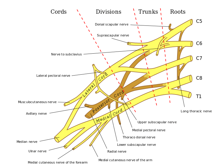
The long thoracic nerve is a pure motor nerve that arises from anterior or ventral roots of C5, C6, and C7 and supplies serratus anterior muscle. Serratus anterior muscle is responsible for stabilization and protraction movement of the scapula.

It is also called posterior thoracic nerve and the external respiratory nerve of Bell, after Sir Charles Bell who provided its first description.

Origin and Course

long thoracic nerve

Long thoracic nerve arises from the anterior rami of three spinal nerve roots: the fifth, sixth, and seventh cervical nerves (C5-C7).

Sometimes, the root from C7 may be absent. Sometimes, there may be a contribution from the C4 root.

The roots from C5 and C6 pierce through the scalenus medius, while the C7 root passes in front of the muscle.

Anatomy of Brachial Plexus

The nerve descends posterior to the roots of the brachial plexus and anterior to the scalenus posterior muscle and has a long course along the chest wall in the mid-axillary line to lie on the superficial surface of the serratus anterior muscle, which it innervates.

The fifth and sixth cervical roots, along with the dorsal scapular nerve, pass through the substance of scalenus medius muscle and join with the root of C7.

The nerve then travels beneath the brachial plexus and clavicle to pass over the first rib. It lies posterior to the first part of the axillary artery.

The nerve descends along the lateral aspect of the chest wall and crosses the outer border of the first rib to reach outer of serratus anterior muscle.

[From its point of appearance to the posterior angle of the second rib, it travels at about  30° posterior angle relative to the anterior axillary line. It then has been noted to pass down between the posterior and middle axillary lines.]

It courses vertically, gets progressively closer to the anterior border of the scapula.

The long thoracic nerve is unique in that it runs superficially to the entire course of the muscle it supplies.

Along with its course over the serratus anterior muscle, the nerve divides into small branches parallel to the main trunk, often several centimeters before turning at right angles and supplying the individual slips by entering the superior aspect of slips.

At the proximal edge of the distal serratus anterior head, it is joined by a collateral branch of the thoracodorsal artery (serratus anterior branch), which crosses over the nerve before dividing into terminal muscle branches. The nerve can be easily identified at this cross-over point, also called Crow’s feet.

Clinical Examination for Long Thoracic Nerve

As the nerve supplies serratus anterior muscle, the examination is done to look for integrity and power of serratus anterior muscle.

The examination involves observation and comparison of both the scapulae and serratus anterior muscles.

Note if there is any the asymmetry between the left and right sides, any atrophy of the muscle, or presence of fasciculations.

Serratus wall test measures and compares the strength of the serratus anterior.

The patient is asked to stand approximately at 2 feet away from the wall, facing it and is asked to push against the wall with the palms flat against the wall at waist level.

Any damage to long thoracic nerve would result in the winging of the scapula due to paralysis of serratus anterior muscle.

Comparison to opposite scapula would help in mild presentations.

Clinical Significance of Long Thoracic nerve

Damage to Long Thoracic Nerve

It may occur for a variety of causes like surgery, trauma to the neck and chest, and sports also result from some sports like archery, tennis etc.

Sports-related nerve damage occurs due to stretching of the nerve. A stretch beyond 10% of the resting length would damage the nerve.

The nerve injury results in serratus anterior paralysis leading to winging of the scapula.

Crow’s foot

It is the point where the long thoracic nerve intersects the most dominant and inferior serratus branch of the thoracodorsal artery

Crow’s foot is an important landmark for flap transfer surgeries.


Related

Spread the Knowledge
48
Shares
 
48
Shares
147   

Filed Under: Anatomy

About Dr Arun Pal Singh

Arun Pal Singh is an orthopedic and trauma surgeon, founder and chief editor of this website. He works in Kanwar Bone and Spine Clinic, Dasuya, Hoshiarpur, Punjab.

This website is an effort to educate and support people and medical personnel on orthopedic issues and musculoskeletal health.

You can follow him on Facebook, Linkedin and Twitter

Primary Sidebar

Browse Articles

Evidence based orthopedics

What is Evidence Based Orthopedics?

Evidence-based orthopedics is the application of principles of evidence-based medicine to the field of orthopedics. Because each of the medical specialty has its own set of challenges and unique applications, therefore, while the core principles remain the same, the methods and the applications must meet the need of each. What is Evidence-Based Medicine and What […]

radiofrequency neurotomy

Radiofrequency Neurotomy – Indications and Procedure

A radiofrequency neurotomy is the use of radiofrequency energy mediated heat to ablate the nerve responsible for facet joint pain or sacroiliac joint pain caused by arthritis or other degenerative changes, or from an injury. Ablation if the nerve beaks the pain signals to the brain, thus eliminating pain. The term radiofrequency ablation is also […]

Popliteal Artery anatomy

Popliteal Artery Anatomy and Course

The popliteal artery is the direct continuation of the superficial femoral artery in the popliteal fossa as the vessel courses posteriorly behind the knee. It gives the main branch as the anterior tibial artery and continues as the tibioperoneal or tibiofibular trunk or posterior tibial artery. It supplies knee, lower leg and foot. Course of […]

Anterior attachments of tibia

Tibia Anatomy and Attachments

Leg is the part of the lower limb between knee and ankle. The osseous framework of the leg is composed of two bones –tibia and fibula. The tibia is a medial and larger of the two bones. The bone is similar to the radius bone of the upper limb. Structure of Tibia Like all bones, […]

Maffucci syndrome has enchondromas and hemangiomas of soft tissue

Maffucci Syndrome Features, Diagnosis and Treatment

Maffucci syndrome is a rare disorder is characterized by enchondromas, bone deformities with venous malformations with or without spindle cell hemangiomas. The condition was first reported by Maffucci in 1881. There is no racial or sexual predilection. It usually manifests early in life, usually around age 4-5 years. About one-fourth of the cases are congenital. But […]

Ectrodactyly in both feet

Ectrodactyly or Lobster Claw Hand (and Foot)

Ectrodactyly is a genetic disorder of skeletal anomaly resulting in the complete or partial absence of fingers or toes due to longitudinal deficiency. Webbing between fingers or toes (syndactyly) may occur giving hands and/or feet a claw-like appearance. The condition has a widely ranging severity and there are combinations of deformities that appear. The malformation […]

radiofrequency neurotomy

Radiofrequency Neurotomy – Indications and Procedure

A radiofrequency neurotomy is the use of radiofrequency energy mediated heat to ablate the nerve responsible for facet joint pain or sacroiliac joint pain caused by arthritis or other degenerative changes, or from an injury. Ablation if the nerve beaks the pain signals to the brain, thus eliminating pain. The term radiofrequency ablation is also […]

© Copyright: BoneAndSpine.com
Manage Cookie Consent
The site uses cookies. Please accept cookies for a better visiting experience.
Functional Always active
The technical storage or access is strictly necessary for the legitimate purpose of enabling the use of a specific service explicitly requested by the subscriber or user, or for the sole purpose of carrying out the transmission of a communication over an electronic communications network.
Preferences
The technical storage or access is necessary for the legitimate purpose of storing preferences that are not requested by the subscriber or user.
Statistics
The technical storage or access that is used exclusively for statistical purposes. The technical storage or access that is used exclusively for anonymous statistical purposes. Without a subpoena, voluntary compliance on the part of your Internet Service Provider, or additional records from a third party, information stored or retrieved for this purpose alone cannot usually be used to identify you.
Marketing
The technical storage or access is required to create user profiles to send advertising, or to track the user on a website or across several websites for similar marketing purposes.
Manage options Manage services Manage vendors Read more about these purposes
View preferences
{title} {title} {title}
 

Loading Comments...