• Skip to primary navigation
  • Skip to main content
  • Skip to primary sidebar
  • Home
  • TeleConsult
  • About
  • Newsletter/Updates
  • Contact Us
  • Policies

Bone and Spine

Orthopedic health, conditions and treatment

  • General Ortho
  • Procedures
  • Spine
  • Upper Limb
  • Lower Limb
  • Pain
  • Trauma
  • Tumors

Shoulder Arthrocentesis Indications and Procedure

By Dr Arun Pal Singh

In this article
    • Indications of Shoulder Arthrocentesis
      • Diagnosis
      • Therapeutic
    • Contraindications
    • Relevant Anatomy
    • Procedure of Shoulder Arthrocentesis
    • Complications
      • Related

Shoulder arthrocentesis is the procedure of aspirating joint fluid from shoulder joint and like other arthrocentesis can have a diagnostic or therapeutic purpose.

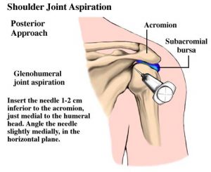
Shoulder joint or the glenohumeral joint can be approached anteriorly or by a posterior approach. The posterior approach allows the patient to be blinded from the procedure, and it mimics the approach used in arthroscopy of the joint.

Anterior approach for shoulder arthrocentesis
Anterior approach for shoulder arthrocentesis, Image Credit: Slideshare

Indications of Shoulder Arthrocentesis

Diagnosis

  • Acute monoarticular [that affects a single joint] arthritis
  • Suspected infection (eg, septic arthritis)
  • Inflammatory joint effusion
    • Gout
    • Pseudogout
    • Rheumatologic disorders
    • Reactive arthropathies

Therapeutic

  • To relieve a large painful effusion.
  • Instillation of medications
  • Repeated arthrocentesis for septic arthritis in carefully selected patients as a means of decreasing bacterial load

Contraindications

  • Cellulitis in the overlying skin
  • Overlying skin lesions (eg, dermatitis or psoriasis)
  • Known bacteremia
  • Bleeding disorders
  • Patient on anticoagulation medication

Relevant Anatomy

The glenohumeral joint of the shoulder is formed by the humeral head and the glenoid fossa of the scapula.

Detailed anatomy of the shoulder joint

Procedure of Shoulder Arthrocentesis

Posterior approach for shoulder arthrocentesis
Posterior approach for shoulder arthrocentesis, Image Credit: http://coxuongkhop.com.vn/images/news/00000609.JPG
  • Shoulder joint can be approached posteriorly or anteriorly. The patient should be seated in a comfortable position.
    • For the anterior approach, the hand of the patient should rest lap so that the shoulder is internally rotated.
    • If the posterior approach is contemplated, rest the patient’s hand on the affected side on the contralateral shoulder.
  • Palpate and mark the needle insertion site according to the approach chosen
  • Anterior approach
    • Palpate the coracoid process and the humeral head.
    • With the arm in internal rotation [internal rotation is achieved by placing the hand in the lap of the patient.], the joint space can be felt as a groove lateral to the coracoid process.
    • Direct the needle slightly laterally and superiorly into the glenohumeral joint space.
  • Posterior approach
    • The needle is inserted 1-2 cm inferior and medial to the posterior tip of the acromion [and directed anteriorly and medially toward the coracoid]
  • Prepare the part according to the entry site chosen. Povidone-iodine and alcohol solution can be used for part preparation. Part preparation is done to sterilize the skin surrounding the insertion site and allow it to dry
  • Administer the local anesthesia using 1% or 2% lidocaine.
  • The joint is aspirated using 18 gauge or thicker needle. Advance the needle while keeping a pull on the plunger of the syringe until synovial fluid is obtained. The amount if the fluid aspirated would depend on the size of the effusion or intended investigation.
  • In case the shoulder is accessed for injection, after aspirating the fluid, stabilize the needle by holding the hub and remove the syringe part and exchange the syringe for a corticosteroid-filled syringe. After the other syringe is in place, the injection can be carried out.
  • Remove the needle after the procedure and apply pressure on the site for few minutes. A small sealant bandage can be applied.
  • Divide the fluid among the specimen tubes and send for the required investigation.

For increased accuracy of needle placement, ultrasound imaging may be used to locate the size of aspiration.

Complications

Complications are few and rare. These include the following.

  • Hematoma
  • Intravascular injection into the axillary vessels
  • Infection is possible
  • Injection of corticosteroids into a tendon or tendon insertion can cause tendon rupture
  • Rise in blood glucose levels in patients with diabetes mellitus in case of corticosteroid instillation.


Related

Spread the Knowledge


Filed Under: Procedures

About Dr Arun Pal Singh

Arun Pal Singh is an orthopedic and trauma surgeon, founder and chief editor of this website. He works in Kanwar Bone and Spine Clinic, Dasuya, Hoshiarpur, Punjab.

This website is an effort to educate and support people and medical personnel on orthopedic issues and musculoskeletal health.

You can follow him on Facebook, Linkedin and Twitter

Primary Sidebar

Browse Articles

anterior knee pain scale

Anterior Knee Pain Scale- Use and Limitations

The anterior knee pain scale is a patient-reported assessment of symptoms and functional limitations in patellofemoral disorders. It was first published in 1993 by Kujala et al. The questions on the Kujala questionnaire add to total of100 points. Anterior knee pain results in significant symptoms and activity limitations. The anterior knee pain scale is a […]

Popliteal Fossa Anatomy

Popliteal Fossa Anatomy and Contents

The popliteal fossa [sometimes called knee pit]  is a diamond-shaped depression lying behind the knee joint, covering the lower part of the femur, and the upper part of the tibia. Boundaries of Popliteal Fossa It becomes easier to understand the landmarks when we visualize looking at the fossa from behind. The boundaries or borders of […]

Foot Sprain

Sprain Causes and Treatment

A sprain is an injury that occurs to ligaments causing stretching or tearing of ligaments. Ligaments are fibrous structures that join two bones across the joints. For example, ankle ligaments connect tibia and fibula bones to the talus and calcaneum across the ankle joint. Sprain originates from French word espriandre which means to wring. Therefore […]

Four Parts of Proximal Humerus Fractures

Proximal Humerus Fracture Causes, Symptoms and Treatment

Proximal humerus fractures account for approximately 5% of all fractures. Proximal humeral fractures generally occur in older patients. This is so because older patients have osteopenic or osteoporotic bones which get damaged easily by trivial trauma. However, high-energy trauma can produce these fractures in any person of any age. Proximal humerus fracture is twice as […]

Exercises for achilles tendon

Exercises For Achilles Tendon Rehabilitation

Achilles tendonitis and Achilles rupture are frequent problems associated with Achilles tendon. Exercises for Achilles tendon are designed for rehabilitation of these conditions. Exercises for Achilles Tendon Rehabilitation after Achilles Tendinosis There are three phases of rehabilitation in Achilles tendinosis In the first and earlier half of the second phase of physical therapy active ankle […]

Suprcondylar Fracture Femur In A Poliotic Limb

Distal Femur Fractures Presentation and Treatment

Distal femur fractures have traditionally been associated with young patients but are increasing in the geriatric population. They are commonly found in young, healthy males and elderly osteopenic females.  With increased use of prostheses,  periprosthetic fractures becoming more common These fractures require anatomically stable fixation. The fractures represent high energy injury in young patients whereas […]

Normal supination of Foot Movement

Supination of Foot and Oversupination

Supination of the foot is the natural movement of the foot as it rolls out during the gait cycle. Supination of foot is the movement of the subtalar joint (between the talus and calcaneus)  and consists of three movements – inversion, plantar flexion, and adduction. Inversion – Sole of the foot is turned inwards Plantar […]

© Copyright: BoneAndSpine.com
Manage Cookie Consent
The site uses cookies. Please accept cookies for a better visiting experience.
Functional Always active
The technical storage or access is strictly necessary for the legitimate purpose of enabling the use of a specific service explicitly requested by the subscriber or user, or for the sole purpose of carrying out the transmission of a communication over an electronic communications network.
Preferences
The technical storage or access is necessary for the legitimate purpose of storing preferences that are not requested by the subscriber or user.
Statistics
The technical storage or access that is used exclusively for statistical purposes. The technical storage or access that is used exclusively for anonymous statistical purposes. Without a subpoena, voluntary compliance on the part of your Internet Service Provider, or additional records from a third party, information stored or retrieved for this purpose alone cannot usually be used to identify you.
Marketing
The technical storage or access is required to create user profiles to send advertising, or to track the user on a website or across several websites for similar marketing purposes.
Manage options Manage services Manage vendors Read more about these purposes
View preferences
{title} {title} {title}
 

Loading Comments...