Last Updated on October 29, 2023
Spinal dysraphism is a group of anomalies where there are malformations in the dorsum of the embryo. There is often an abnormal fusion of the midline embryonic neural, vertebral and mesenchymal structures.
The estimated incidence of spinal dysraphism is about 1–3 per 1000 live births.
The incidence of the spinal dysraphism is declining in the world over in the last few decades due to the better nutrition for women, folic acid supplementation and improved antenatal care.
There is a multifactorial causation, involving both genetic and environmental.
Pathology of Spinal Dysraphism
Spinal dysraphism can be broadly divided into two different pathological entities
- open spinal dysraphism
- meningocele
- myelomeningocele
- Occult or closed spinal dysraphism
- dorsal dermal sinus
- lipomyelomeningcele
- diastematomyelia
- neurenteric cyst
- thickened filum terminale
Tethered cord syndrome is the term used for occult dysraphism when the symptoms arise due to a tethered cord which can force the spinal cord to stretch as they grow leading to progressive spinal cord damage if untreated.
The tethered cord syndrome is a clinical diagnosis based on signs and symptoms.
TCS is causally linked to Chiari malformation and any diagnosis of tethered cord syndrome must be followed by screening for Chiari’s several degrees.
Ehlers-Danlos syndrome or Klippel-Feil syndrome should also be screened for.
Assessment of Newborn
Examination of the head and neck involves the assessment of
- Head size and shape
- Skull bones, and openness of fontanelles
- Lacunar skull defects and the size of the posterior fossa
- Level of the lesion
- The condition of the skin
- The extent of the skin defect
- Associated deformities
- Deformities of lower limb
- Neurological examination
Clinical Presentation
Open dysraphism
It presents with a swelling over the back which is noticed at birth.
The symptoms are due to CSF leak or the exposed spinal cord.
The skin over the swelling may give way during labor, resulting in CSF leak, contamination, and meningitis.
Defects predominantly involve thoracolumbar, lumbosacral, lumbar, thoracic, sacral and cervical region in decreasing order of frequency.
Older children present with back pain and/or neural deficits which include motor, sensory and sphincter dysfunction, depending upon the severity and level. [Leg weakness or gait abnormalities and urinary irregularities like incontinence or retention.]
Chiari malformation presents with lower brainstem and lower cranial nerve dysfunction.
The associated skeletal abnormalities are kyphosis, scoliosis, and deformities of the long bones and feet, hemivertebrae, defective ribs, etc.
Swelling, hairy patches, dimples, or fatty tumors on the lower back may be found.
In case of occult spinal dysraphism, the tethered spinal cord syndrome may go undiagnosed until adulthood, when neurological deficits start occurring. These relate to the degree of strain on the spinal cord over time.
Tethering may also develop after spinal cord injury.
Syringomyelia may occur due to blockage of fluid by scar tissue and cyst formation due to fluid pressure.
This can lead to additional loss of movement or feeling, or the onset of pain or autonomic nervous system symptoms.
In adults, the onset of symptoms typically includes –
- Severe pain in the back that radiates to the legs, groin, and perineum
- Motor and sensory deficit in lower limbs
- Loss of feeling and movement in lower extremities
- Urinary incontinence or retention
- Progressive neuropathic bladder
- Urinary frequency and urgency
- The feeling of incomplete voiding
- Poor voluntary control
- Urge and stress incontinence
- Chronic recurrent urinary infections
- Renal stones
- Renal failure
- Bowel control problems
Neurological symptoms can include a mixed picture of upper and lower motor neuron findings.
Prenatal Diagnosis of Spinal Dysraphism
It is done by
- Ultrasound
- May identify up to 90% cases of myelomeningoceles
- Maternal Alpha Feto Protein screening
- Performed around 12, 22 and 32 weeks.
- Can detect spina bifida [80%] and anencephaly [90%]
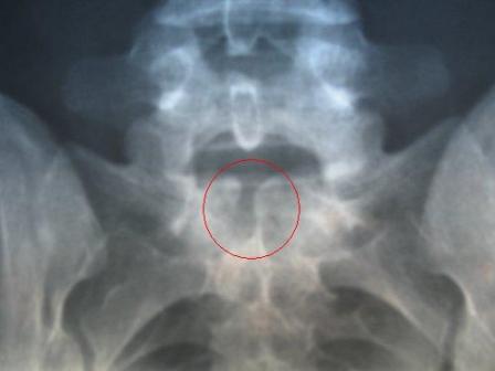
Imaging
Plain X-rays
X-rays reveal the skull defects, spine deformities, and bony anomalies.
MRI
MRI is the investigation of choice to study the neural tissue abnormalities and also to assess the severity of hydrocephalus and Chiari malformation. It is also considered to be the gold standard for diagnosing a tethered cord.
Ultrasonography
It can be used to obtain to assess hydrocephalus, a tethered cord in children less than 6.
A tethered cord is often diagnosed as a “low conus.”
[The conus medullaris normally terminates at or above the L1-2 disc space. Therefore a conus below the L1-2 disc space may indicate a tethered cord.]
Management of Spinal Dysraphism
Treatment is mostly surgical. Surgery should be undertaken as soon as it is practical.
In case of suspected meningitis or CSF infection or colonization of the wound, prophylactic antibiotics and anticonvulsants should be given.
The newborn child with myelomeningocele should have saline dressings till surgery.
In adults, the surgical treatment is detethering or spine-shortening vertebral osteotomy.
Detethering involves removal of structures that tether the cord.
Vertebral osteotomy aims to indirectly relieve the excess tension on the spinal cord by removing a portion of the spine. This procedure offers a unique benefit in that the spinal cord remains fixated to the spine, preventing retethering and spinal cord injury as possible surgical complications.
Symptomatic and supportive treatment includes Medications such as NSAIDs, opiates, synthetic opiates, COX-2 inhibitors, and off-label applications of tricyclic antidepressants combined with anti-seizure drugs.
Some Important Pathologies
Spina Bifida
Spina bifida is discussed here.
Lipomyelomeningocele
Lumbosacral lipomyelomeningocele is a subcutaneous fibrofatty mass that traverses the lumbodorsal fascia, causing a spinal laminar defect, penetrating dura and tethering of the spinal cord.
Presence of lipomatous mass or a cutaneous marker is diagnostic in 90% of the children.
The surgical treatment is aimed at complete untethering and prevention of retethering of the cord.
Dermal sinus
Congenital dermal sinuses are a unique form of occult dysraphism presenting with meningitis, tethering or neurological compression
The dermal sinus tracts are lined by squamous epithelium and may penetrate anywhere in the midline from the lumbosacral region to the occiput or nasion. Dermoid and epidermoid nodules are frequently associated with these tracts.
The neurological examination is nearly always normal. The MRI is diagnostic.
There are usually in the midline, providing a portal for infection leading to meningitis or intraspinal abscesses.
Neurological examination except in cord compression. The treatment includes antibiotics and complete surgical excision of the sinus, tract and the associated lesions.
Occult Tight Filum Terminale
The filum terminale thickens or hardens and causes downward pressure on the spinal cord. This can actually cause scoliosis as well as most of the other symptoms of the more typical tethered cord.
References
- Steinbok P. Dysraphic lesions of the cervical spinal cord. Neurosurg Clin N Am. 1995;6:367–76
- Gupta RK, Sharma A, Jena A, Tyagi G, Prakash B, Khushu S. Magnetic resonance evaluation of spinal dysraphism in children. Childs Nerv Syst. 1990;6:161–5.
- Boop FA, Russell A, Chadduck WN. Diagnosis and management of the tethered cord syndrome. J Ark Med Soc. 1992;89:328–31
- Lhowe D, Ehrlich MG, Chapman PH, Zaleske DJ. Congenital intraspinal lipomas: Clinical presentation and response to treatment. J Pediatr Orthop. 1987;7:531–7.
- Kanev PM, Park TS. Dermoids and dermal sinus tracts of the spine. Neurosurg Clin N Am. 1995;6:359–66.
- Caruso R, Cervoni L, Fiorenza F, Vitale AM, Salvati M. Occult dysraphism in adulthood. A series of 24 cases. J Neurosurg Sci. 1996;40:221–5.