Last Updated on March 12, 2020
The thoracic cage or rib cage is the skeletal framework that encloses the contents of the thorax.
The thoracic cage or rib cage along with its contents form the thorax portion of the body. Roughly speaking, this is the area of the chest.
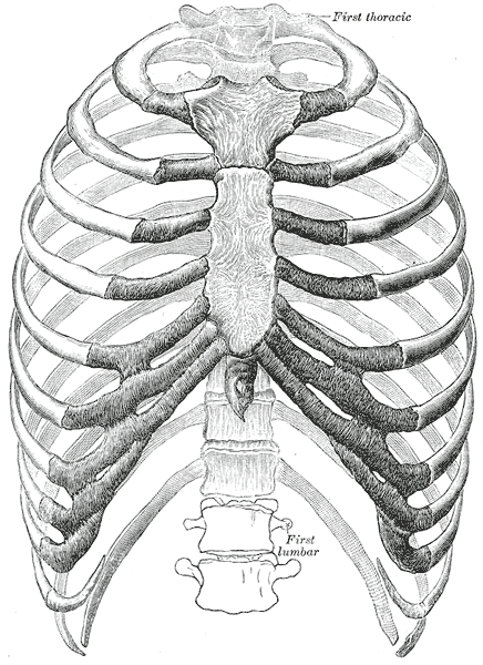
The Thoracic cage consists of the 12 pairs of ribs with their costal cartilages and the sternum.
The ribs are uniquely arranged to maximize the thoracic cavity volume and anchor posteriorly to the 12 thoracic vertebrae (T1–T12).
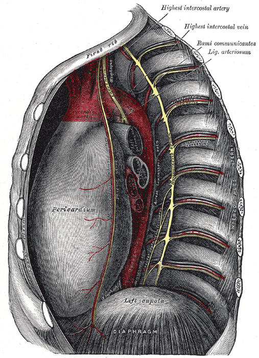
The thorax contains organs of respiration [lungs] and circulation [heart]. The functioning of both of these is very vital for life. The thoracic cage protects the heart and lungs and assists the functions of respiration and circulation by increasing and decreasing thoracic cavity volumes.
Structure of Thoracic Cage
Thoracic cage is formed by bones and cartilage [osseocartilaginous]. It is an elastic cage which is primarily designed for increasing and decreasing the intrathoracic pressure so that air is sucked into the lungs during inspiration and expelled during expiration.
Thoracic cage is formed anteriorly by the sternum, posteriorly by the 12 thoracic vertebrae and the intervening intervertebral discs and on each side. The ribs on both the sides complete the cage.
Each rib articulates posteriorly with the vertebral column.
The thorax resembles a truncated cone which is narrow above and broad below. The narrow upper end is continuous with the root of the neck from which it is partly separated by the suprapleural membrane or Sibson’s fascia [a covering on the lung].
The broad lower end is almost completely separated from the abdomen by the diaphragm.
In the transverse section, the thorax is bean-shaped or kidney-shaped except in infants below the age of two years, where it is circular.
Individual elements of the thoracic cage are discussed further.
Sternum
The sternum is the anterior anchor of the thoracic cage. It an elongated and fat bone that consists of the following part
- Manubrium
- Body
- Xiphoid process
The manubrium is the wide superior part of the sternum. It has a shallow, U-shaped upper border called the jugular or suprasternal notch that can be easily palpated at the neck base, between two medial ends of clavicle.
On either side at the superolateral margin, the manubrium has a notch for clavicle. It is called clavicular notch and the site of the sternoclavicular joint
The first ribs also attach to the manubrium on either site.

The body of the sternum refers to its elongated central part of the sternum. The manubrium and body join together at an angle, called the sternal angle
The second rib attaches to the sternum at the sternal angle.
This rib is the first to be palpated on examination as the first rib is behind the clavicle.
The sternal angle and second rib are important landmarks for the counting of ribs.
The sternal body gives attachment to third to seventh ribs.

The xiphoid process is the inferior tip of the sternum. It is a cartilaginous structure in early life but becomes ossified gradually with aging.
Ribs
There are a total of 12 pairs of ribs.
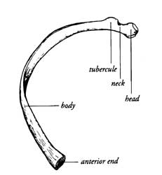
Each rib is a curved, flattened bone that articulates posteriorly with the thoracic vertebrae and attaches anteriorly to the sternum via their costal cartilages.
Beginning at the top, the ribs are numbered 1–12 in accordance with their articulation with the thoracic vertebrae.
Though ribs are bony structures, anteriorly, each rib ends in costal cartilage which varies in its dimensions for different ribs. This costal cartilage then attaches to the sternum.
There are two main types of ribs – true ribs and false ribs depending on articulation with the sternum.
- True ribs are ribs which articulate with sternum via individual costal cartilage. Each of these ribs articulates with vertebra on the posterior side and sternum on the anterior side via costal cartilage These are also called vertebrosternal ribs. First 7 ribs are true ribs
- False ribs do not articulate with sternum. Anteriorly, these join the costal cartilage of a superior rib. Such ribs are called vertebrochondral ribs. Ribs 8-10 are false ribs
- Floating ribs are false ribs that do not attach to sternum at all and disappear in the muscles of the lateral abdominal wall. These are also called vertebral ribs. 11th and 12th ribs are floating
The costal cartilages of the 7th, 8th, 9th and 10th ribs form the costal margin.
The anterior ends of the 11th and 12th ribs are free and these ribs are termed as floating ribs. Some authors also call them vertebral ribs.
The Head of the rib forms the posterior end of a typical rib and articulates with the costal facet located on the body of the same numbered thoracic vertebra and that of the next higher vertebra.
The neck of the rib is narrowed part and anatomically in a lateral position to the head.
Tubercle of the rib is a small bump on the posterior surface of the rib and articulates with the facet on the transverse process of the same numbered vertebra.
Beyond neck, as we move anteriorly is the shaft of the rib.
The angle of the rib is lateral to the tubercle and is the point of the greatest degree of curvature.
The angles of the ribs form the most posterior limit of the thoracic cage and are in line with the medial border of the scapula.
Inferior margin of the rib has a groove for the passage of blood vessels and nerve.
In infants the ribs are horizontal. Therefore the respiration is only by action of the diaphragm.
In adults the thorax is oval. The ribs are oblique and their movements alternately increase and decrease the diameters of the thorax. This results in the drawing in of air into the thorax (inspiration) and its expulsion (expiration). This is called thoracic respiration.
Adults have both diaphragmatic and thoracic respiration.
Joints of Thorax
Manubriosternal Joint
- Between manubrium and sternum
- Secondary cartilaginous joint.
- Only slight movements of the body of the sternum on the manubrium during respiration.
- Fused in 10%
Costovertebral Joints
Costovertebral joint is between the head of a typical rib and two vertebrae to form two-plane synovial joints. One vertebra is of the same level as that of rib whereas the other is the body of the next higher vertebra
Costotransverse Joints
Costotransverse joint is a joint between the tubercle of a typical rib and the transverse process of the corresponding vertebra. It has the following ligaments
- Ligaments
- Capsular ligament
- three costotransverse ligaments
- Superior costotransverse ligament
- Extends from the crest on the neck of the rib to the transverse process of the vertebra above
- has two laminae
- Inferior costotransverse ligament
- From the posterior surface of the neck to the transverse process of its own vertebra.
- Lateral costotransverse ligament
- Connects the lateral nonarticular part of the tubercle to the tip of the transverse process.
Chondrosternal Joints
The first chondrosternal joint is a primary cartilaginous joint. It does not permit any movement. This helps in the stability of the shoulder girdle and of the upper limb.
The 2nd to 7th costal cartilages articulate with the sternum by synovial joints. Each joint has a single cavity except in the second joint where the cavity is divided into two parts. The joints of the thorax are held together by the capsular and radiate ligaments.
Costochondral Joints
The cartilaginous joints between the sternal end of ribs and the lateral ends of costal cartilages.
Each rib has a depression shaped like a cup that the costal cartilage articulates with.
There is normally no movement at these joints of the thorax.
Interchondral Joints
The 5th to 9th costal cartilages articulate with one another by synovial joints. The tenth cartilage is united to the ninth by fibrous tissue.
Muscles of Thorax
Muscles of thoracic age are the intercostals (external, internal and innermost), subcostals, and transversus thoracis.
All these muscles function to change the volume of the thoracic cavity during respiration.
Some other muscles that are not part of the thoracic wall but attach to it are the pectoralis major, minor, serratus anterior and the scalene muscles.
Intercostals
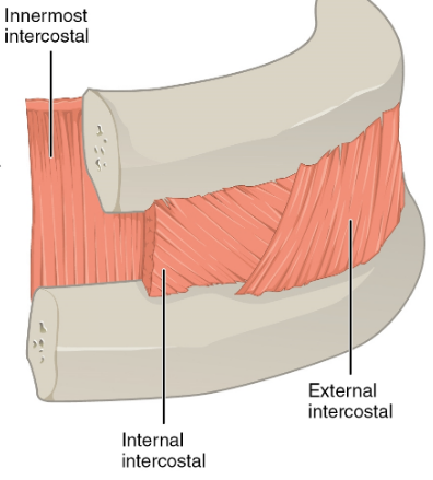
These muscles are found in the intercoastal spaces i.e. the spaces between the ribs. These are three types of muscles and are arranged in three layers.
All the intercoastal muscles are supplied by intercostal nerves [T1- T11].
External Intercostals
- 11 pairs
- Run inferoanteriorly from the lower border rib above to the upper border of the rib below
- Continuous with the external oblique of the abdomen.
- Elevates the ribs to increase the thoracic volume.
Internal Intercostals
- Deep to the external intercostals
- Run the same extent as external intercostals but in opposite direction (inferoposteriorly). From the lateral edge of the costal groove and inserts into the superior surface of the lower rib.
- Continuous with the internal oblique muscle of the abdominal wall.
- Interosseous part decreases the thoracic volume by depressing the ribs
- The interchondral part elevates the ribs.
Innermost Intercostals
- Deepest intercostal muscles
- Similar structure to the internal intercostals
- Originates from the medial edge of the costal groove and inserts into the superior surface of the lower rib.
- Separated from the internal intercostals by the intercostal neurovascular bundle
- Action similar to internal
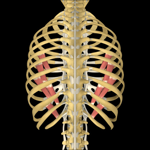
Transversus Thoracis
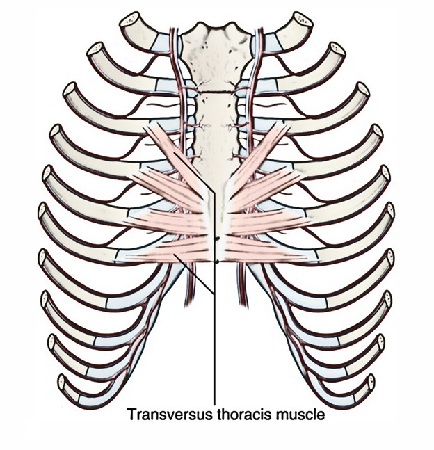
- Extends from the posterior surface of the inferior sternum to the internal surface of costal cartilages
- Continuous with transversus abdominis inferiorly.
- Weak depressor of the ribs
- Supplied by intercostal nerves (T2-T6).
Subcostals
- Found in the inferior portion of the thoracic wall
- Extends from the inferior surface of the lower ribs, near the angle of the rib to the superior border of the 1-2 ribs below
- The fibers are parallel to that of the innermost intercostals
- Action is similar to the action of the internal intercostals
- Supplied by intercostal nerves