Last Updated on October 11, 2020
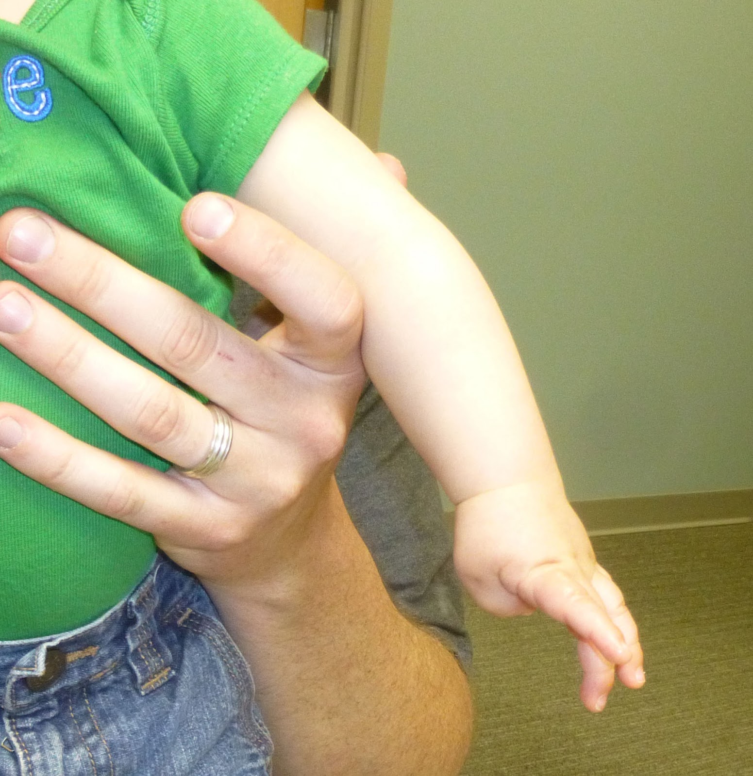
The congenital clasped thumb is a rare condition where the thumb is positioned in adduction and extreme flexion at the metacarpophalangeal joint and interphalangeal joints.
The term implies a spectrum of thumb anomalies that range from mild thumb extension limitation to severe abnormalities of the thenar muscles, web space, and soft tissues.
The condition is also called congenital contracted or clenched thumb.
The deformity occurs due to an imbalance between the flexors and extensor systems of the thumb.
The diagnosis is made after observation for 3 months and if no active extension at the metacarpophalangeal joint is shown after a prolonged observation. This is because in early life the natural placement of thumb is similar and resolves by 3 months of age.
The anomaly is almost always bilateral and males are more commonly affected than females.
The familial incidence has been observed.
Causes, Risk Factors, and Pathophysiology
The exact factors causing the condition are not known. risk factors
Exact causative factors are not well known
The following are the probable pre-disposing factors-
- Marriages in blood relation [Consanguineous marriages]
- Family history
The condition is transmitted as autosomal dominance inheritance but the expression is variable.
Structurally, there is attenuation or deficiency of extensor pollicis brevis or extensor pollicis longus or both.
There may be associated contracture of
- First web space
- Adductor pollicis
- First dorsal interosseous muscle
There could also be global instability of the first metacarpophalangeal joint and abnormality of the articular cartilage of the first metacarpophalangeal joint.
There are few abnormalities of the limbs which may be present along with this condition. These are
- Flexion deformities of the four fingers of hand
- Congenital vertical talus
- Congenital talipes equinovarus
- Often bilateral
The conditions which are associated with clasped knife thumb are
- Arthrogryposis
- Digitotalar dysmorphisms [a type of arthrogryposis]
- Freeman-Sheldon syndrome [Multiple contractures in the body]
- X-linked MASA syndrome [spastic paraplegia]
Classification of Congenital Clasped Thumb
[McCarroll and Tsuyuguchi’s]
- Type I
- Thumb supple
- Extensor mechanism absent or hypoplastic
- Thumb can be passively abducted and extended a
- No other digital anomaly present.
- Type II
- Clasped thumb with joint contractures
- Thumb cannot be passively extended and abducted.
- Other digital anomalies may be present
- Collateral ligament abnormality
- First web space contracture
- Abnormality of thenar muscles.
- Type III
- Rigid clasped thumb
- Associated with arthrogryposis or other similar syndromes
- Minimal extensor mechanism abnormality
Clinical Presentation
The complaint is about the thumb deformity where the child does not extend his thumb and keep it pressed against the palm.
The deformity is usually on both sides.
It needs to be noted that for about the initial three months of infancy the thumb grasp reflex is normal. In this, the thumb is flexed across the palm and the fingers are flexed over the clutched thumb. But on stimulation and even spontaneously, the infant actively extend his fingers and thumb.
This is an important feature to differentiate.
Therefore, the diagnosis of a congenital clasped thumb is made with certainty after 3 months of age.
On examination, there would be flexion-adduction deformity of the thumb [and exists beyond 3rd month of life and affects both the sides.
When contracture is present, there may be associated with pain.
Hypoplasia of the extensor pollicis brevis, extensor pollicis longus, and abductor pollicis longus may be present resulting in various positions of thumb.
Associated musculoskeletal and other deformities should be evaluated.
Another entity that congenital clasped thumb can be confused with is Congenital Trigger thumb deformity.
Trigger thumb is a flexion deformity of the thumb where the root cause is entrapment is present at A1 pulley level due to fusiform enlargement of the flexor pollicis longus tendon.
Presence of bilateral deformity, presence of associated conditions, and congenital lack of extensor mechanism point towards the clasped thumb.
The presence of a nodule in the thumb [Notta’s node] is suggestive of a trigger thumb.
Imaging
Xrays of bilateral hands and upper limbs should be done to find any bony abnormality and associated syndromes.
Treatment of Congenital Clasped Thumb
The treatment of congenitally clasped thumb depends on the following factors
- Disease stage
- Age at presentation,
- Other pathologies
Type I and some type II cases generally show improvement with conservative methods.
In type III and those type II cases which do not improve with conservative treatment, surgical treatment produces good results.
Across all the stages, conservative treatment is not advised for the cases who have agenesis or severe hypoplasia of extensor pollicis brevis.
Nonoperative Treatment
This consists of serial splinting and stretching for 3-6 months and should be first line treatment for all the types of congenitally clasped thumb. The treatment should be started around the age of 6 months.
Nonoperative treatment produces good results in type I deformities when one the tendons, extensor pollicis longus or brevis are present.
But results are poor results when both the tendons are absent.
Operative Treatment
The surgery is indicated in
- Type II and III cases
- Type I cases with absent tendons
- Those who respond poorly to conservative treatment
Various surgical options are
Extensor Indicis Prporius Tendon Transfer to EPL
This procedure is indicated in type I or II with residual deficiency in active extension
Thumb reconstruction
The procedure is done in following indications
- Failed conservative treatment
- Type III deformity with first web space soft tissue deficiency
- Type II or III deformity with significant metacarpophalangeal joint contractures
The procedure is done after 3years fo age and involves
- Widening and of the first space
- Skin flap transfers
- Transposition flap of skin (dorsal rotational advancement flap)
- Release of the origins of thenar muscles from transverse carpal ligament
- Tendon transfers
- Z lengthening of flexor pollicis longus
Arthrodesis
This option is used in severe deformities when skin release and tendon trasnfer cannot help.
References
- Medina J, Lorea P, Marcos A, et al. Flexion deformities of the thumb: clasped thumb and trigger thumb. Chir Main 2008; 27:35–39.
- Chen CC, Shieh SJ, Chiu HY. Re: subluxation of extensor pollicis longus and brevis complex as a cause of congenital clasped thumb. J Hand Surg Eur 2009; 34:268–269.
- Mih AD. Congenital clasped thumb. Hand Clin 1998; 14:77–8]
- Ghani HA, El-Naggar A, Hegazy M, et al. Characteristics of patients with congenital clasped thumb: a prospective study of 40 patients with the results of treatment. J Child Orthop 2007; 1:313–322.