Last Updated on November 22, 2023
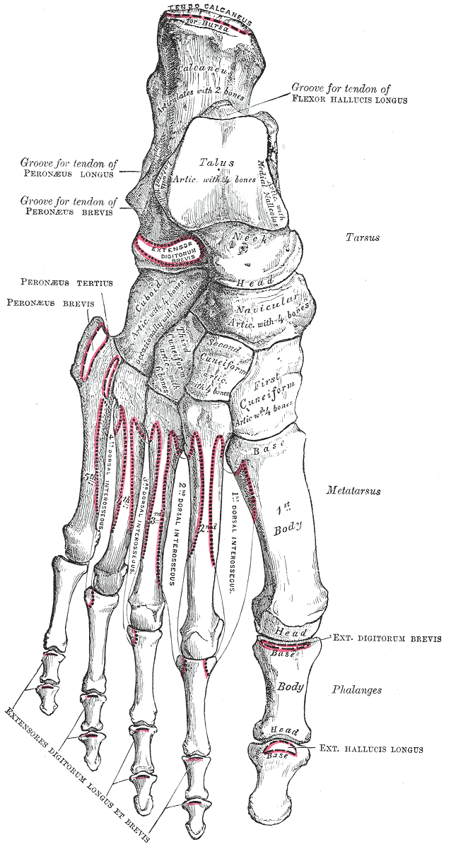
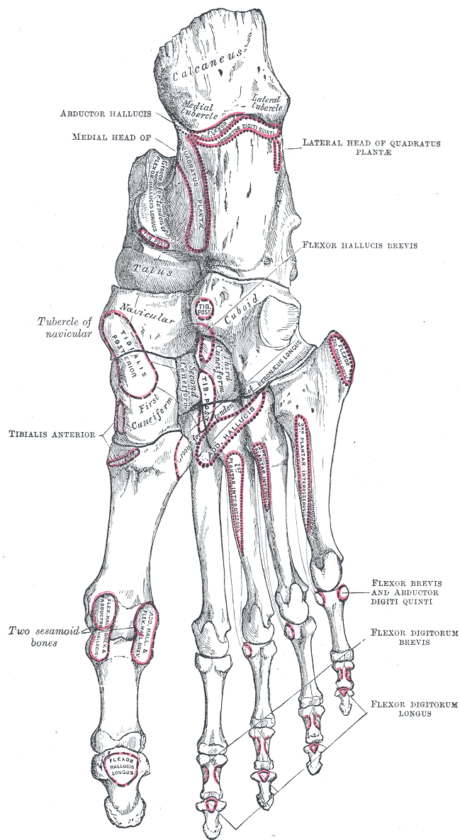
There are seven tarsal bones. The proximal row is formed by the talus above, and the calcaneus below. The distal row contains, from medial to lateral side, medial cuneiform, the intermediate cuneiform, the lateral cuneiform and the cuboid.
Navicular is interposed between the talus and the three cuneiform bones. In other words, it is interposed between the proximal and distal rows.
Each tarsal bone is roughly cuboidal in shape, having six surfaces.
Calcaneus and Talus
Calcaneus and talus tarsal bones are discussed individually.
Cuboid
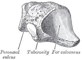
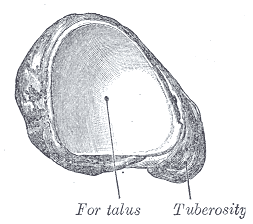
The cuboid is the lateral most bone of the distal row. It is situated in front of the calcaneum and behind the 4th and 5th metatarsal bones. It has six surfaces.
It is cuboidal in shape but has a broader base oriented medially

Structure of Cuboid Bone
- Dorsal surface
- Rough for the attachment of ligaments.
- Directed upwards and laterally.
- Plantar surface
- Crossed anteriorly by an oblique groove [peroneal sulcus] bounded posteriorly by a prominent ridge
- The groove runs obliquely anteromedially and lodges the tendon of the peroneus longus.
- The ridge ends laterally in an eminence, the tuberosity
- Tuberosity presents an oval facet for gliding of sesamoid bone or cartilage frequently found in the tendon of the peroneus longus.
- Lateral surface
- Short and notched.
- Medial surface
- Partly articular and partly nonarticular
- Oval facet in the middle articulates with the lateral cuneiform bone.
- Proximal to this a small facet may be present for the navicular bone.
- Rough in the rest of its extent, for the attachment of strong interosseous ligaments.
- Proximal or posterior surface
- Concavoconvex for articulation with the anterior surface of the calcaneum.
- Inferomedial angle projects backward as a process, which underlies and supports the anterior end of the calcaneus.
- Distal surface
- Triangular and articular
- Divided by a vertical ridge into two areas. The medial, quadrilateral in form, articulates with the fourth metatarsal
The cuboid articulates with
- Calcaneus
- Third cuneiform
- Fourth metatarsal
- Fifth metatarsal
Note* – It occasionally articulates with a fifth, the navicular.
Attachments on Cuboid
- The notch on the lateral surface and the groove on the plantar surface
- Occupied by the tendon of the peroneus longus.
- Ridge posterior to the groove
- Attachment of deep fibers of the long plantar ligament.
- Rough surface behind the groove
- Plantar calcaneocuboid ligament
- Few fibers of the flexor hallucis brevis
- A fasciculus from the tendon of the tibialis posterior.
- Plantar Surface
- Posterior Border – The short plantar ligament
- The posteromedial part
- A slip from the tibials posterior
- Origin to the flexor hallucis brevis.
- Medial surface
- Ligaments including the lateral limb of the bifurcate ligament.
Ossification of Cuboid
The cuboid bone ossifies from one centre which appears just before birth.
Navicular
The navicular bone is boat-shaped. It is situated on the medial side of the foot, in front of the head of the talus, and behind the three cuneiform bones.
It forms the uppermost portion of the medial longitudinal arch of the foot and acts as a keystone of the arch.
The navicular bone has 6 surfaces.
- Posterior or proximal surface
- Oval, concave, broader laterally
- Articulates with the rounded head of the talus.
- Medial surface
- Has a blunt and prominent tuberosity, directed downwards
- The tuberosity is separated from the plantar surface by a groove
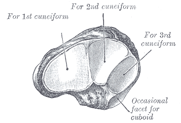
- Anterior surface
- Convex
- Divided into 3 facets for the three cuneiform bones.
- Dorsal surface
- Convex from side to side
- Rough for the attachment of ligaments
- Plantar surface
- Irregular and rough for the attachment of ligaments.
- Lateral surface
- Rough and irregular for the attachment of ligaments
- Occasionally presents a small facet for articulation with the cuboid bone.
Attachments on Navicular
- Tuberosity of the navicular bone
- Tibialis posterior
- Groove below the tuberosity transmits a part of the tendon of this muscle to other bones.
- Plantar surface
- Attachment to the spring ligament.
- Lateral Surface
- Calcaneonavicular part of the bifurcate ligament
- Dorsal Surface
- Talonavicular, cuneonavicular and cubonavicular ligaments
Ossification of Navicular
Navicular ossifies from one centre which appears during the third year of life.
Articulations of Navicular
The navicular articulates with 4 bones: the talus and the 3 cuneiforms. Sometimes, a fifth articulation for the cuboid is also present.
Cuneiform Bones
There are three cuneiform bones, medial, intermediate or middle and lateral.
The medial cuneiform is the largest and the intermediate or middle cuneiform, the smallest.
Cuneiforms are wedge-shaped bones. In the medial cuneiform, the edge of the wedge forms the dorsal surface. In the intermediate and lateral cuneiforms, the thin edge of the wedge forms the plantar surface.
The medial and lateral cuneiforms project farther distally than the middle cuneiform to create a mortise for the base of the second metatarsal that articulates with the middle cuneiform.
See the image below

Medial Cuneiform
It is the largest cuneiform bone. It is situated at the medial side of the foot, between the navicular behind and the base of the first metatarsal in front. It has following surfaces
- Medial surface
- Broad, quadrilateral and subcutaneous.
- Anterior inferior edge carries a smooth oval impression [for part of tibialis anterior tendon]
- Rest of surface is rough
- Lateral surface
- Marked by an inverted L-shaped facet
- Along the posterior and superior margins
- For the intermediate cuneiform bone
- Anterosuperior part of the facet is separated by a vertical ridge
- Ridge creates an anterior area for articulation with the base of the second metatarsal bone
- The anteroinferior part of the lateral surface is rough for the attachment of ligaments and part of the tendon of the peroneus longus.
- Marked by an inverted L-shaped facet
- Anterior surface
- Convex
- Much larger than the posterior surface
- Articulates with the first metatarsal bone.
- Posterior surface
- Triangular and concave
- Articulates with the most medial and largest of the 3 facets on the anterior surface of the navicular [piriform facet].
- Plantar surface
- Formed by the base of the wedge
- Rough posterior part has a tuberosity for the insertion of fibers of the tendon of the tibialis posterior.
- Anterior part provides insertion to part of the tendon of the tibialis anterior.
- Dorsal surface
- Narrow end of the wedge
- Directed upward and lateralward
- Rough for the attachment of ligaments.
The first cuneiform articulates with
- Navicular
- Second cuneiform
- First metatarsal
- Second metatarsal.
Attachments of First Cuneiform
- The greater part of the tibialis anterior is inserted into an impression on the anteroinferior angel of the medial surface.
- The plantar surface receives a slip from the tibialis posterior.
- A part of the peroneus longus is inserted into the rough anteroinferior part of the lateral surface.
Middle Cuneiform
It is the smallest of three cuneiforms. It forms a dorsal base and apex is plantar.
Situated between the other two cuneiforms, it articulates with the navicular behind and the second metatarsal in front.
- Anterior surface
- Triangular and narrower than the posterior
- Articulates with the base of the second metatarsal bone.
- Posterior surface
- Also triangular
- Articulates with the middle facet on the anterior surface of the navicular.
- Medial surface
- L-shaped articular facet, along the superior and posterior borders, for articulation with the medial cuneiform
- Rest of medial surface is rough for the attachment of ligaments.
- Lateral surface
- Has a smooth facet for articulation with the third cuneiform bone.
- Dorsal surface
- Forms the base of the wedge.
- Plantar surface
- Sharp and tuberculated
- Rough for the attachment of ligaments and for the insertion of a slip from the tendon of the tibialis posterior.
The medial cuneiform articulates with
- Navicular
- First cuneiform
- Third cuneiform
- Second metatarsal.
Attachments
The plantar surface receives a slip from the tibialis posterior.
Lateral Cuneiform
- Dorsal surface
- Oblong
- Formed by the base of the wedge
- Its posterolateral angle prolong backward
- Plantar surface
- Formed by the edge of the wedge
- Has a rounded margin
- Attachments
- Part of the tendon of the tibialis posterior
- Part of the flexor hallucis brevis
- Ligaments.
- Posterior or proximal surface
- Rough in its lower one third
- Has a triangular facet in its upper two thirds for the navicular bone.
- Lateral surface
- Marked in its posterosuperior part by a triangular or oval facet for the cuboid
- At anteroinferior angle, a small facet may be present for the fourth metatarsal bone.
- Anterior surface
- Triangular articular facet
- Articulates with the third metatarsal bone.
- Rough, nonarticular area serves for the attachment of an interosseous ligament.
- The 3 facets for articulation with the 3 metatarsal bones are continuous with one another
- The facet for articulation with the cuboid is usually separate.
- Triangular articular facet
- Anterior surface
- Triangular articular facet
- Articulates with the third metatarsal bone.
- Rough, nonarticular area serves for the attachment of an interosseous ligament.
- Medial surface
- marked along its posterior margin by a vertical strip indented in the middle, for articulation with the intermediate cuneiform bone
- Along the anterior margin of the surface there is a facet, sometimes divided, for the base of the second metatarsal bone
The third cuneiform articulates with
- Navicular
- Intermediate or middle cuneiform
- Cuboid
- Second, third, and fourth metatarsals.
Ossification of Cuneiforms
Each cuneiform bone ossifies from one center, which appears
- Lateral cuneiform – first year in the lateral cuneiform
- Medial cuneiform – second year
- Intermediate cuneiform – third year