Last Updated on May 21, 2022
Spinal shock is a state of total sensory, motor power loss, and loss of all reflexes for a period after spinal cord injury [complete or near-complete injury]. It is followed by the gradual recovery of reflexes distal to injury with often residual neurological deficits.
[Read more about spinal cord injury]
It begins within a few minutes of the spinal injury, it may take several hours before the full effects occur. It generally occurs in acute sudden injury but is also seen in injury that develops over few hours.
The nervous system is unable to transmit signals from the brain to end organs as they are not routed by the spinal cord.
Spinal shock is defined as a state of transient physiologic (rather than anatomic) reflex depression of cord function below the level of injury, with associated loss of all sensorimotor functions.
In contrast to other kinds of shocks [hemorrhagic shock, neurogenic shock], spinal shock does not mean circulatory collapse but rather a state of depressed spinal reflexes distal to spinal cord injury.
Usually, the spinal shock recovers within 24 hours but may last over a few weeks in less common cases. In some rare cases, spinal cord shock can last for several more months.
The term Spinal Shock was created by Hall in 1840 and later the definition was modified by Sherrington.
Pathophysiology of Spinal Shock
The exact cause of the spinal shock is not known. It is thought that acute injury causes depolarization of axons due to the transfer of kinetic energy.
Experiments have shown the progression of spinal cord injury as
- Hemorrhage in grey matter
- Protein accumulation in grey matter
- Edema then occurs [peaks at 3-6 days]
- Slow central cord necrosis and vacuolization for two months
Primary injury occurs due to the initial impact with underlying chronic cord compression. These impacts are
- Fracture-dislocation
- Burst fractures
- Rupture of disc
- Direct laceration due to bone or metal
There are multiple mechanisms suggested for secondary injury [vascular, inflammation, ionic channels, free radicals, etc. ] but molecular mechanisms of secondary injury remain unknown.
After the injury, the transection of the cord causes spinal shock. These are the main suggested mechanisms
- 1. Synaptic changes in cord segments below injury
- Increase in presynaptic inhibition [suppression of neurotransmitter release]
- High concentration of glycine [inhibitory neurotransmitter]
- Hyperpolarization of spinal motor neurons [will not respond to stimulus]
- Sudden withdrawal of facilitatory inputs from the brain
- Loss of normal function in spinal pathways and their interconnections
There are three phases of this
Phase 1
- Complete loss or weakening of all reflexes below the level of spinal cord injury
- Lasts for about a day
- The neurons involved in various reflex arcs become hyperpolarized and less responsive to brain inputs
Phase 2
It occurs over the next two days and is characterized by the return of some, but not all, reflexes. The superficial reflexes such as the palntar reflex, bulbocavernosus reflex recover first.
Monosynaptic reflexes, such as the deep tendon reflexes, are not restored until Phase 3.
The reason reflexes return is the hypersensitivity of reflex muscles following denervation — more receptors for neurotransmitters are expressed and are therefore they are easier to stimulate.
Phases 3 and 4
- Hyperreflexia, or abnormally strong reflexes
- Reflex results from minimal stimulation
- It occurs following the sprouting of interneurons and lower motor neurons below the injury [to attempt to the reestablishment of neural connections]
Causes
Any instance that causes spinal injury will invariably cause spinal shock. Here are the most common causes of spinal injury
- Motor vehicle accidents
- Most common cause
- Falls
- Mechanical injury from tumor or abscess
In addition to the primary happenings above, further injury to the spinal cord may occur due to disruption of blood supply by the primary event. The resultant decrease in blood supply and damage to the cord due to lack of oxygen further damage the spinal cord.
Transient spinal shock has been reported with the use of intrathecal iodinated contrast.
Risk Factors
The presence of certain risk factors makes one prone to spinal cord injury
- Cervical spondylosis
- Most common
- Present in about 10%
- Congenital abnormalities of the spine
- Aatlantoaxial instability
- Congenital fusions
- Tethered Cord
How to Identify Spinal Shock in a Spinal Injury Patient
Spinal shock is clinically manifested as
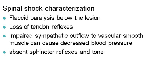
- Loss of spinal cord function below the level of the injury
- Flaccid paralysis
- Loss of sensation
- Absent bowel and bladder control
- Loss of reflexes
In addition to the spinal shock, a patient of a motor vehicle accident with a spinal injury may have neurogenic or hemorrhagic shocks. These must be assessed and managed.
Patients should be examined for other concomitant injuries to rule out critical injuries first. All spinal injuries are considered unstable till it has been ruled out. So proper spinal injuries precaustions must be taken.
Neural examination must be done as per ASIA scale. ASIA stands for the American Spinal Injury Association Scale, and it helps to to quantify the neurological deficit resulting from a spinal cord injury.
A complete spinal injury or spinal shock have similar findings. However, in case of incomplete spinal injury too the the presence of spinal shock would present it as complete injury. It is only after spinal shock is over, the spared neural functions can be ascertained if not involved by the initial injury.
Thus, clinically, whether the injury is complete or incomplete can only be told when spinal shock goes away.
Findings in case of complete spinal injury or spinal shock are
- No sensation in the levels below the injury
- No motor power below the injury – flaccid paralysis
- Absent reflexes
- Bowel and bladder incontinence
- Bradycardia and decreased blood pressure due autonomic dysregulation
Thus There is total paralysis, hypotonia & areflexia, and at its conclusion, there may be hyperreflexia, hypertonicity, and clonus.
Recovery from Spinal Shock
The recovery occurs from distal to proximal [caudorostral pattern].
The duration of the shock is variable. Superficial reflexes may recover within hours whereas deep tendon reflexes may take months even.
The recovery of plantar reflex is considered now as the earliest sign that marks the end of spinal shock. The reflexes come back in the following order
- Delayed plantar reflex
- Bulbocavernosus reflex
- Cremasteric reflex
- Ankle jerk
- Babinski sign
- Knee jerk
Earlier teachings considered return of reflex activity below the level of injury (such as bulbocavernosus) as an end of the spinal shock.
Return of the of plantar and bulbocavernosus reflex signifies the end of the spinal shock, and if the injury is complete, any further neurological improvement will be minimal. In such cases, it is highly unlikely that significant neurologic recovery will occur.
Spinal shock does not occur in the lesions that occur below the cord, and therefore, lower lumbar injuries should not cause spinal shock. If bulbocavernosus reflex is absent in such cases may indicate a cauda equina injury
Delayed Plantar Reflex
The plantar reflex is a reflex where the toes flex when the foot is stimulated with a blunt instrument. This reflex is normally present in healthy adults.
An opposite movement, the toe extension is known as Babinski response or Babinski sign and is seen in upper motor neuron lesions or as a primitive reflex in newborns.
Bulbocavernous Reflex
Bulbocavernosus reflex can be checked by noting anal sphincter contraction in response to squeezing the glans penis or tugging on the Foley catheter that would be in place due to bladder incontinence. It involves the S1, S2, S3 nerve roots and is a spinal cord-mediated reflex. Its presence signals the end of the spinal shock.
The significance of Spinal Shock
There is a loss of signal transmission and the loss of these signals will result in loss of movements, sensations other body functions. Complete loss of movement and sensation below the level of the spinal cord injury makes it difficult to assess the exact quantum of injury. Thus it is not possible to find the level, extent, and severity of the injury as patients would show a complete neural loss.
The only way to find that is to wait for the spinal shock to recover. Over the first few weeks, the systems adjust to the effects of the injury and their function improves. Therefore, during this time it is unlikely that an accurate prediction of any recovery or permanent paralysis can be made.
Management of Spinal Shock
This essentially involves the management of spinal cord injury and is discussed in detail in the concerned article.
Here only brief points are given to create an outline
- management of hypotension and bradycardia
- Aim mean arterial blood pressure around 90 mmHg
- Fluids
- Drugs – Norepinephrine, dopamine or phenylephrine, Midodrine, desmopressin, atropine
- Management of paralytic ileus if present otherwise enteral feeding
- Venous thromboembolism prophylaxis, as
- Urinary catheterization i
- Intravenous steroids – methylprednisolone
- Nursing care for the prevention of ulcers and contractures
- Definitive treatment of the injury/Rehabilitation
References
- Atkinson PP, Atkinson JL. Spinal shock. Mayo Clin Proc. 1996 Apr;71(4):384-9. [Link]
- Boland RA, Lin CS, Engel S, Kiernan MC. Adaptation of motor function after spinal cord injury: novel insights into spinal shock. Brain. 2011 Feb;134(Pt 2):495-505. [Link]
