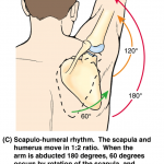
Scapulohumeral rhythm is the interplay between the scapula and the humerus during motion of the shoulder. There is a definitive pattern of interaction of scapula and humerus when contributing to the motion of the shoulder. Scapulohumeral rhythm is also called glenohumeral rhythm. Optimal function of the shoulder is reliant on the coordinated movement of the […]
Anatomy
Lumbosacral Plexus Anatomy
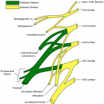
Lumbosacral plexus is formed anterior or ventral rami of L1–S3 roots. All major nerves of the lower extremity are derived from lumbosacral plexus. Though functionally considered as one entity, the lumbosacral plexus is usually thought of anatomically as an upper lumbar plexus and a sacral plexus also called lower lumbosacral plexus [also receives lumbar contribution]. […]
Pudendal Nerve Anatomy and Function
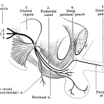
The pudendal nerve is a major somatic nerve of the sacral plexus originating from S2-S4 nerve roots, innervates the external genitalia of both sexes and the skin around the anus, anal canal and perineum and supplying pelvic muscles, the external urethral sphincter and the external anal sphincter. It also carried carries sympathetic nerve fibers to […]
Transitional Vertebra Definition and Types
A transitional vertebra at any junction is characterized by features retained from two adjacent regions in the vertebral column. For example in the lumbosacral transitional vertebra, there is an L5 vertebra that can become like S1 vertebra or S1 can leave the sacrum and become an L6. As we know from our knowledge of the […]
Lumbosacral Transitional Vertebra or LSTV
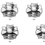
Lumbosacral transitional vertebra refers to a spectrum of are congenital spinal anomalies involving L5 and adjacent vertebrae and varies from complete fusion of L5 with the sacrum, called sacralization to complete separation of S1 from the sacrum and present as L6, called lumbarization. When the L5 vertebra fuses completely to the sacrum, 4 lumbar vertebrae […]
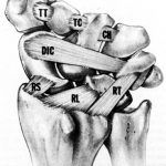
Scaphoid Bone Anatomy
The scaphoid bone is one of the carpal bones of the wrist on the lateral side of the wrist. Carpal bones are arranged in two rows and scaphoid is most lateral of the proximal row. The carpus is made up of 8 carpal bones, which are arranged in two rows. Proximal row It contains (from […]
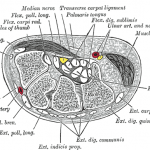
Wrist Anatomy- Bones, Joints, Muscles and Ligaments
The wrist is a complex joint formed by a collection of multiple bones and joints bridges the hand to the forearm. It is an ellipsoidal joint. The bones comprising the wrist include Distal ends of the radius and ulna 8 carpal bones Proximal portions of the 5 metacarpal bones Wrist carries complex articulations that allow […]
Normal Alignment of Lower Limb – Axes and Orientation

To understand the deformities of the lower limb, it is important to grasp and establish the parameters defining normal alignment. The normal alignment of lower limb is governed by the arrangement of the femur, tibia, hip, knee, and ankle. To understand it better, the complex three-dimensional shapes of bones and joints can be simplified to basic […]
Blumensaat Line and Its Significance
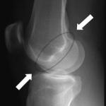
Blumensaat line is a line which corresponds to the roof of the intercondylar fossa of the femur as seen on a lateral radiograph of the knee joint. It is drawn on lateral projection x-ray of the knee. Blumensaat line is important because the angle that it forms on the radiograph with various other lines can be used to determine the position of the […]
Meniscofemoral ligaments – Humphery and Wisberg Ligaments
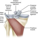
Meniscofemoral ligaments are straight bands of collagen that attach to the posterior horn of lateral meniscus and lateral part of medial femoral condyle. While some consider them one ligament with two bands others consider them as two distinct ligaments. These ligaments are named based on their location in relation to the posterior cruciate ligament. The […]