• Skip to primary navigation
  • Skip to main content
  • Skip to primary sidebar
bone and spine logo

Bone and Spine

Your Trusted Resource for Orthopedic Health Information

  • Home
  • About
  • Contact Us
  • Policies
  • Show Search
Hide Search
You are here: Home / Sports Injuries and Rehabilitation / A Case of Medial Malleolus Fracture Fixed By Percutaneous Fixation

A Case of Medial Malleolus Fracture Fixed By Percutaneous Fixation

Dr Arun Pal Singh ·

Last Updated on March 15, 2020

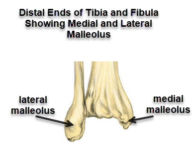
Medial malleolus fracture is a fracture of the distal medial projection of tibia bone called medial malleolus. Medial malleolus and lateral malleolus contribute forms the socket of the ankle joint where talus articulates. There are three malleoli in the ankle. The medial and the posterior arise from the tibia.

A counterpart called lateral malleolus is the distal end of the fibula.

medial malleolus and lateral malleolus
Medial malleolus and lateral malleolus, Posterior malleolus not visible being on the backside [posterior]
A fracture is a break in the continuity of the bone and is usually caused by forces  beyond the strength of the bone.

Medial malleolus fracture is a common ankle injury.

Actually, fractures of medial malleolus are part of the spectrum of malleolar fractures and another ankle injuries. It rarely occurs in isolation and is often accompanied by a fracture of lateral malleolus [most common] or posterior malleolus or ligament injury.

Following is an example of undisplaced medial malleolus fracture.

Xray Ankle showing fracture of medial malleolus


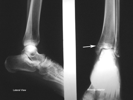
The x-ray shows a fractured medial malleolus.

Like other fractures, treatment depends upon many factors that are injury-related and patient-related. These are the presence of a concomitant fracture of the fibula, amount of displacement of fracture fragments, the age of the patient and demands and expectations of the patient.

The treatment of the medial malleolus fracture can be nonoperative or operative.

Nonoperative treatment or conservative treatment consists of immobilization in below-knee or short leg casts. In some cases, where initial injury has led to substantial swelling, the patient may be given a temporary splint first.

After splinting, the swelling is allowed to reduce before a definitive cast is given.

Nonoperative treatment is mostly done in cases of undisplaced fractures or patients with very low functional demands.

Surgical treatment of medial malleolus fracture aims at anatomical restoration and holding the fractured fragments at that anatomical position by means of screws, K-wires or tension band wiring.

A fractured medial malleolus would take around 4-6 weeks to unite.

Fractures of medial malleolus have a very good prognosis when these are treated appropriately.

Contents hide
1 A case of Percutaneous Fixation of Medial Malleolus Fracture
2 Further Study

A case of Percutaneous Fixation of Medial Malleolus Fracture

The patient had a history of twisting of ankle and broke medial malleolus. Following is the anterior and posterior x-rays of ankle showing the medial malleolus x-ray.

Preoperative x-ray showing vertical fracture of medial malleolus
Preoperative x-ray showing a vertical fracture of medial malleolus

Being an active adult patient and considering the configuration of the fracture, it was decided that the fracture would be fixed. As the fracture was minimally displaced, an attempt to fix the fracture percutaneously without disturbing the soft tissues was made.

The patient was investigated and pre-anesthetic evaluation done.

Spinal anesthesia was used and the patient was kept in a supine position. The limb was externally rotated for better access from the medial side.

The fracture was checked in Carm.

The fracture was held in position with the help of K-wire jockey stick.

The hole for the first screw was drilled after confirming the position in C-arm.

After drilling the hole a malleolar screw was inserted. Here is the preoperative x-ray of the screw being inserted.

C-arm image shows the first screw is being inserted after drilling. The fracture was held in position by by a Kwire before screw was inserted
C-arm image shows the first screw is being inserted after drilling. The fracture was held in position by a K-wire before screw was inserted

Similarly, the second screw was inserted.

Second screw hole point being confirmed under C-arm

After the second screw was inserted, the positions were checked.

Lateral view after insertion of both screws
Confirmatory AP view after insertion of both screws
AP view after insertion of both screws


The incision sites for screw insertion were stapled.

Post operative clinical photograph showing minimal incision
Postoperative clinical photograph showing the minimal incision

The immediate postoperative x-ray showed a well-reduced fracture in both AP and lateral views.

Postoperative AP and lateral x-rays of ankle showing well reduced fracture and satisfactory screw replacement
Postoperative AP and lateral x-rays of ankle showing a well-reduced fracture and satisfactory screw placement

The patient was followed up over a period.

Staples were removed after 12 days.

The fracture showed good union on serial x-rays.

Weight-bearing was allowed after 8 weeks of surgery.

Here is an x-ray done after 3 months of surgery.

X-rays 3 months after surgery showing good union
X-rays 3 months after surgery showing good union of medial malleolus fracture

The fracture is well united and the ankle is well reduced.

Further Study

  • Malleolar Fractures of Ankle- Bimalleolar Fractures, Trimalleolar Fractures

Save

Sports Injuries and Rehabilitation This article has been medically reviewed by Dr. Arun Pal Singh, MBBS, MS (Orthopedics)

About Dr Arun Pal Singh

Dr. Arun Pal Singh is a practicing orthopedic surgeon with over 20 years of clinical experience in orthopedic surgery, specializing in trauma care, fracture management, and spine disorders.

BoneAndSpine.com is dedicated to providing structured, detailed, and clinically grounded orthopedic knowledge for medical students, healthcare professionals, patients and serious learners.
All the content is well researched, written by medical expert and regularly updated.

Read more....

Primary Sidebar

Know Your Author

Dr. Arun Pal Singh is an orthopedic surgeon with over 20 years of experience in trauma and spine care. He founded Bone & Spine to simplify medical knowledge for patients and professionals alike. Read More…

Explore Articles

Anatomy Anatomy Fractures Fractures Diseases Diseases Spine Disorders Spine Disorders Patient Guides Patient Guides Procedures Procedures
featured image of gower sign for segmenatal instability of lumbar spine

Clinical Tests for Lumbar Segmental Instability

Lumbar segmental instability may not always be visible on standard …

mesurement of scoliosis for braces

Braces for Scoliosis- Types, Uses and Results

Braces for scoliosis are recommended to prevent the scoliotic curve …

discogenic back pain

Discogenic Back Pain Causes, Diagnosis and Treatment

Discogenic back pain is a common cause of axial low back pain [the …

Elbow arthrodesis using internal fixation

Elbow Arthrodesis- Indications, Methods and Complications

Elbow arthrodesis refers to the surgical fusion of the elbow joint. It …

skeletal traction in upper tibial pin

Skeletal Traction – Indication, Uses and Complications

Skeletal traction is a type of traction where the force is applied …

Popular articles

Anatomy of Calcaneus

Calcaneus Fracture – Causes, Presentation and Treatment

Calcaneus fracture or os calcis …

erythromelalgia images

Erythromelalgia Causes and Treatment

Erythromelalgia is a rare disorder …

Degrees of freedom Knee

Normal Biomechanics of Knee and Movements

Knee allows locomotion with minimum …

spina bifida

Spina Bifida- Causes, Types and Management

Spina bifida is a  neural tube defect …

Bone and Spine

© 2025 BoneAndSpine.com · All Rights Reserved
The content provided on BoneAndSpine.com is intended for informational and educational purposes only. It is not a substitute for professional medical advice, diagnosis, or treatment. Read Disclaimer in detail.